Fluorescence Transitions
White light and fluorescence – before/after images
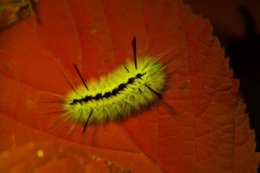
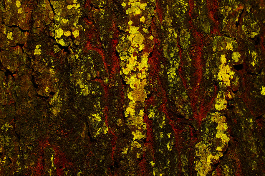
Enjoy the magical transformation of fluorescence in this selection of paired images.
Just put your mouse over an image and move left/right to control the slider – no need to click.
Science Labs
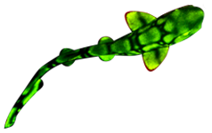
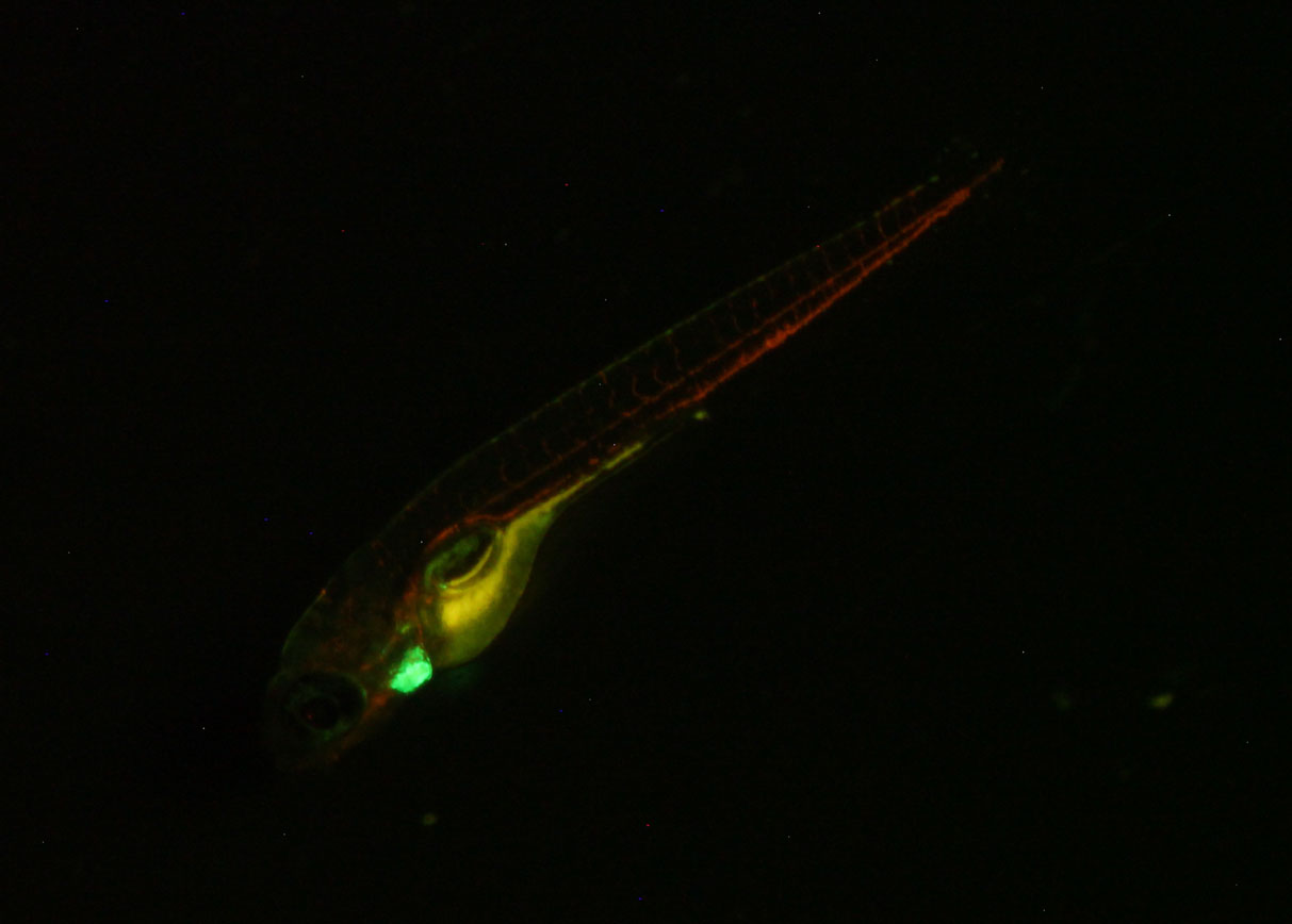
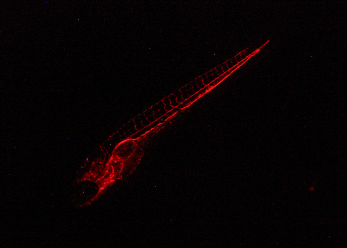
Zebrafish expressing cmlc:GFP (heart) and gata1:dsRed (blood cells)
Royal Blue excitation shows the strong green heart fluorescence, with weaker expression from the blood cells. The yellow is autofluorescence from the yolk.
Green excitation brings out the red fluorescence in the blood cells.
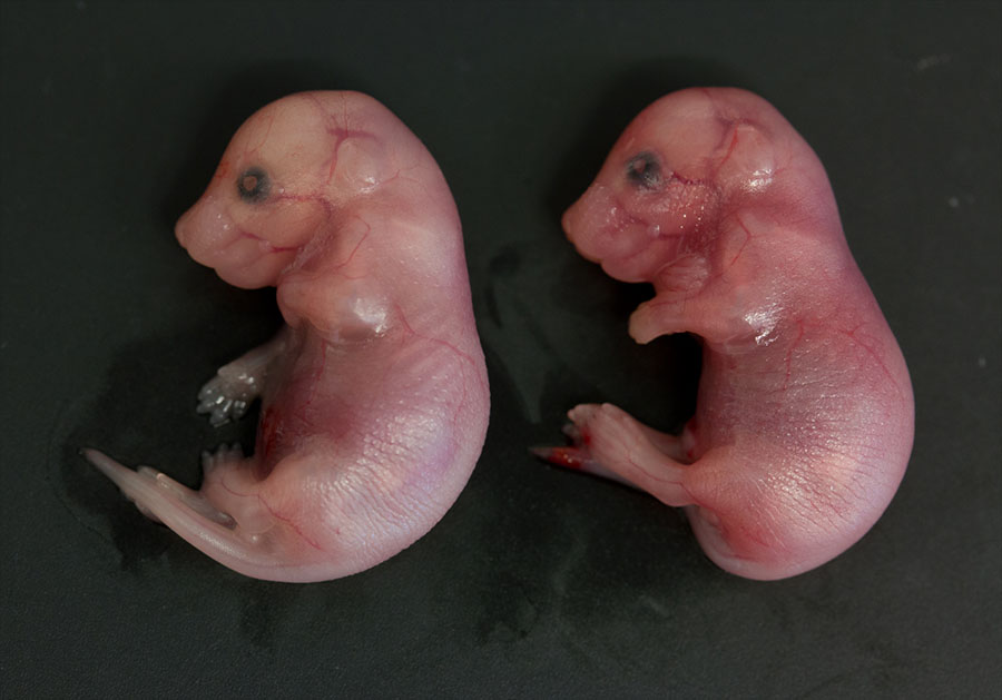
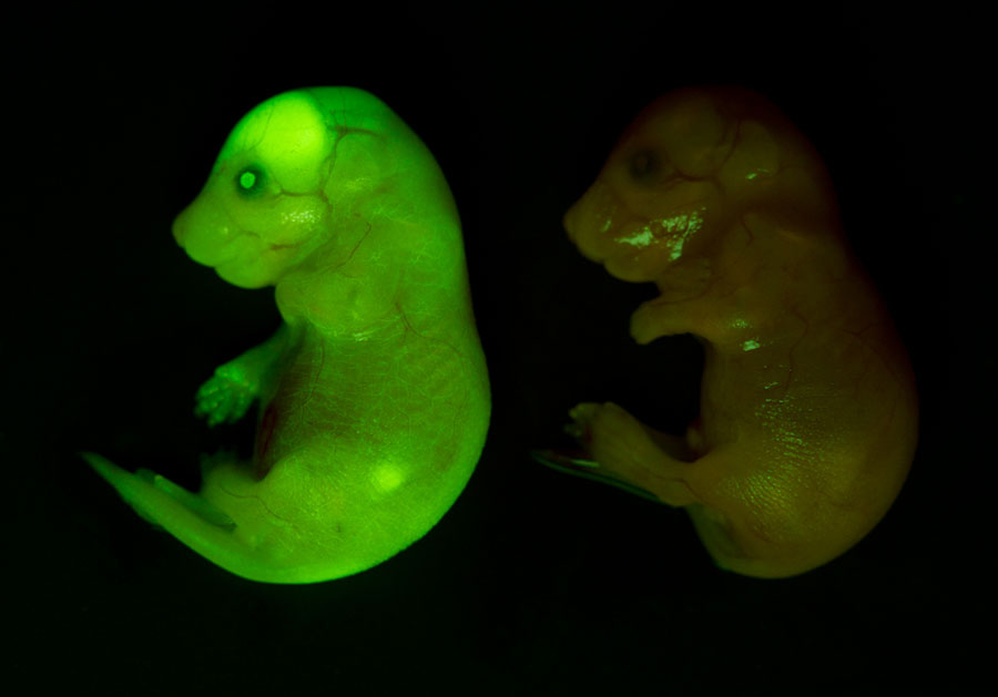
Mouse pups – one GFP-positive, the other not
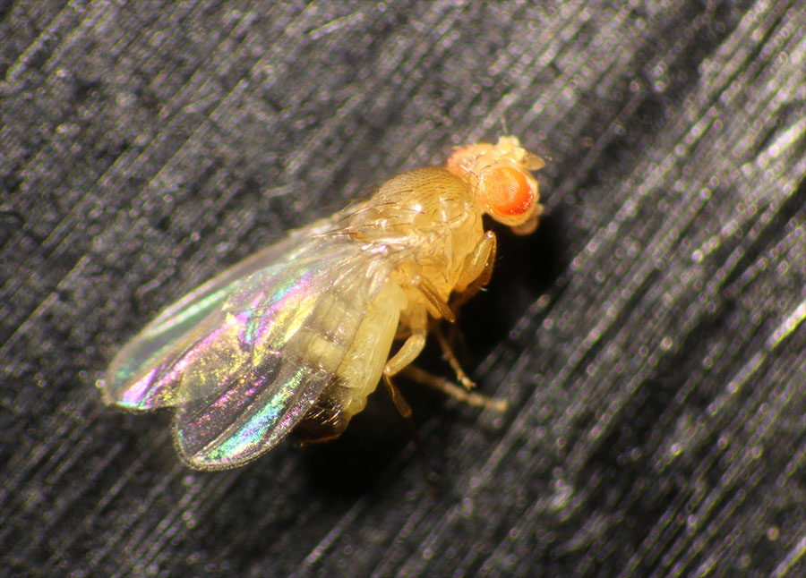
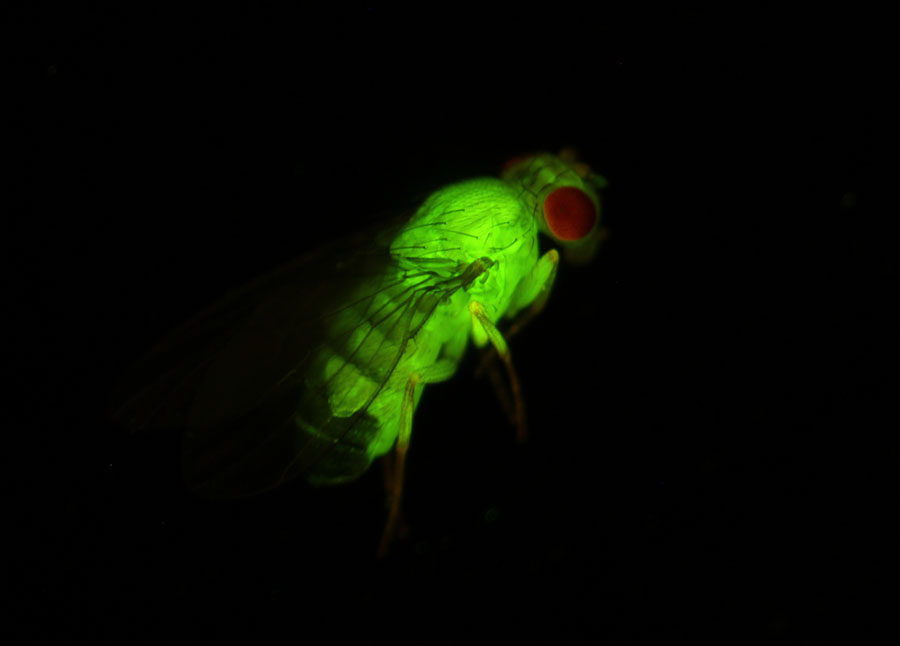
Drosophila expressing GFP
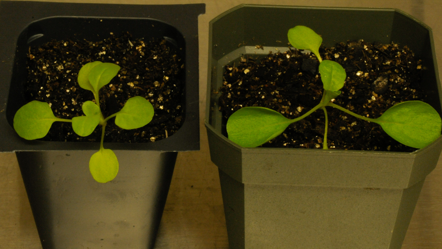
Crop engineering research – RFP-positive and -negative seedlings

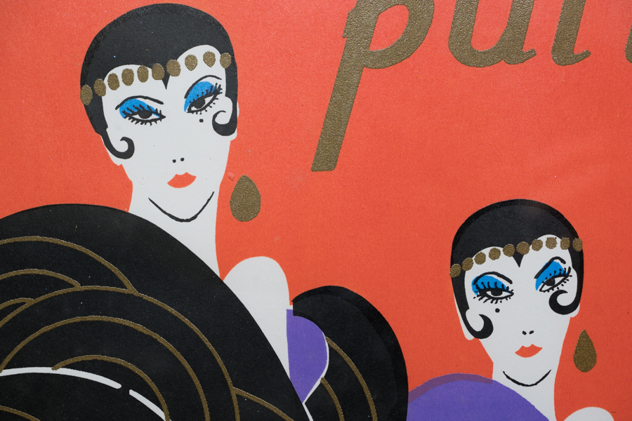
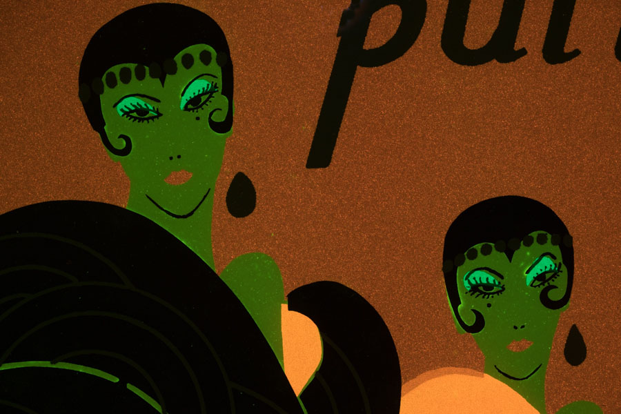
Museum and Art Research and Conservation
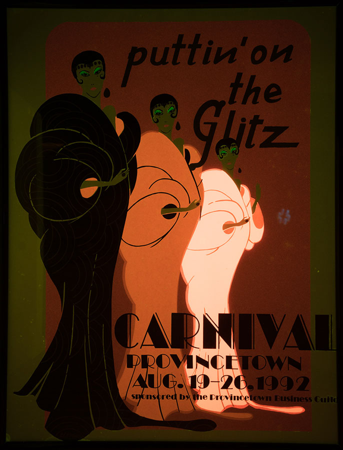
Poster print

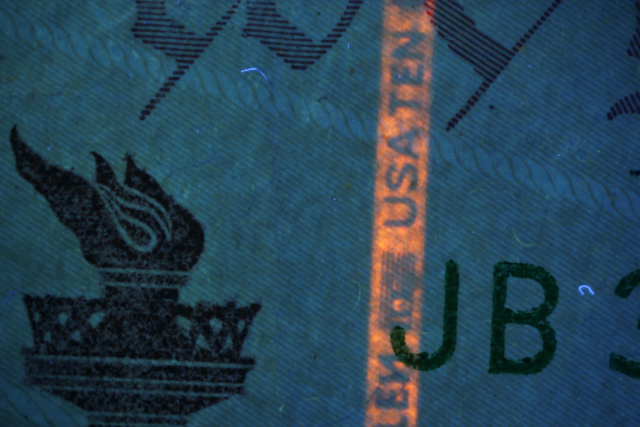
Forensic Sciences
Anti-counterfeit fluorescence stripe on a US$10 bill

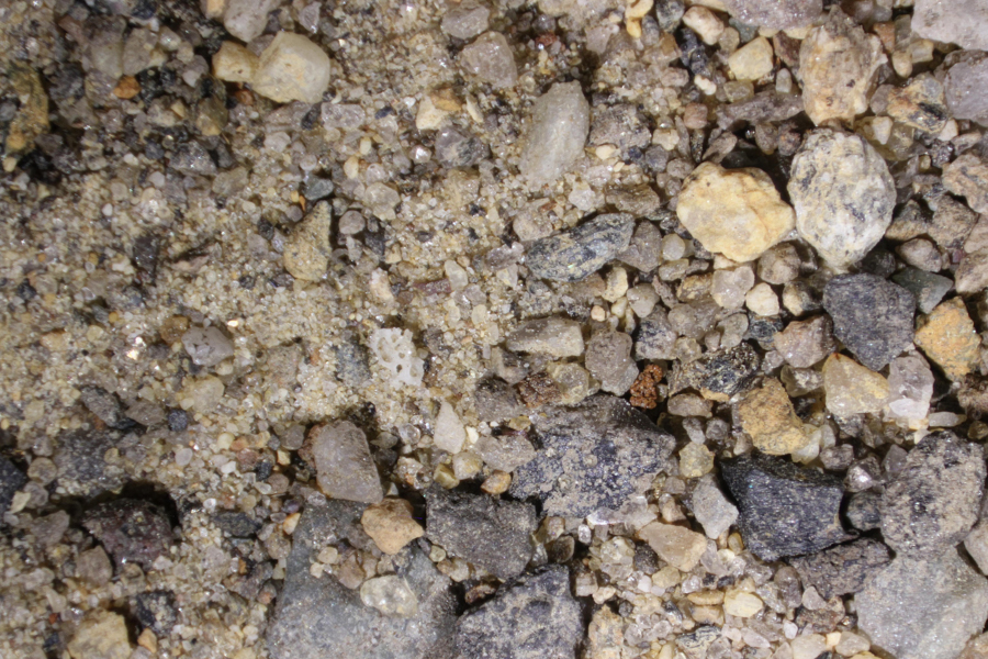
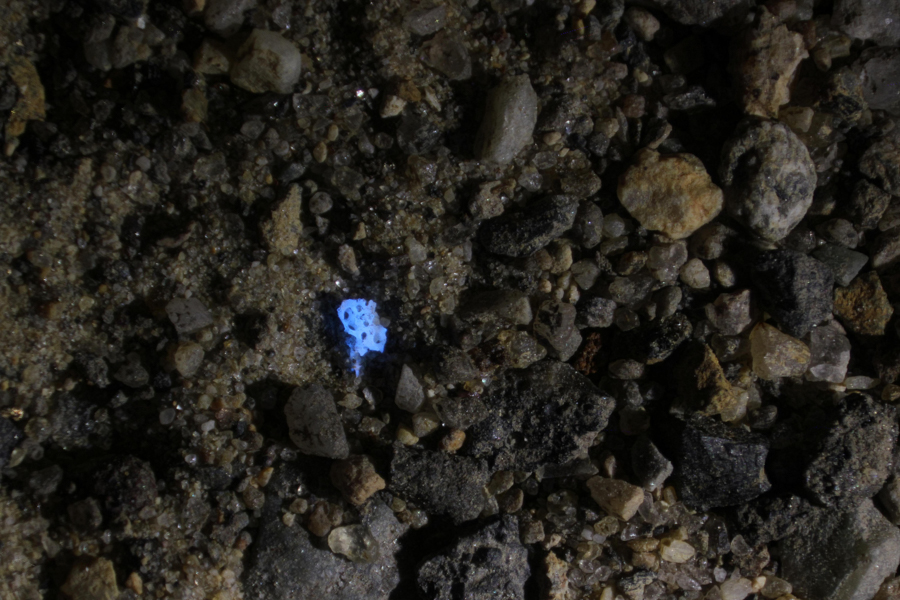
Bone fragment in dirt
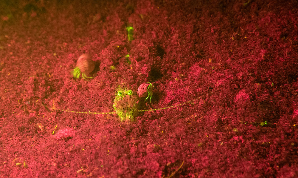
Marine Life – Science & Sport
Shrimp, New England
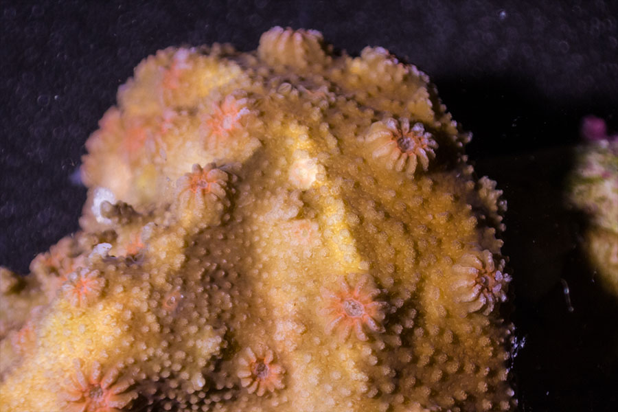
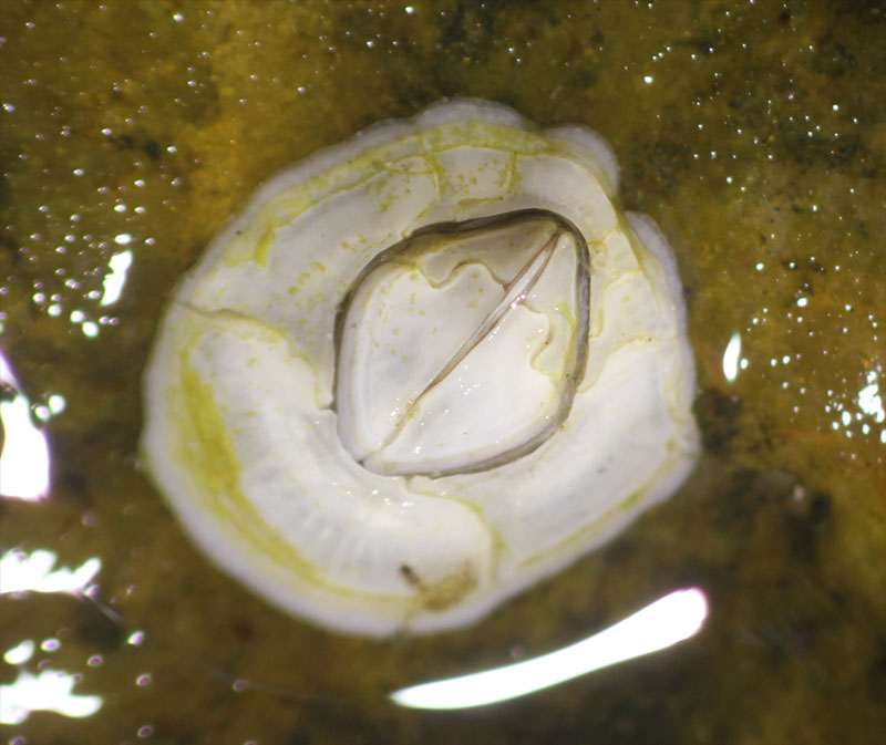
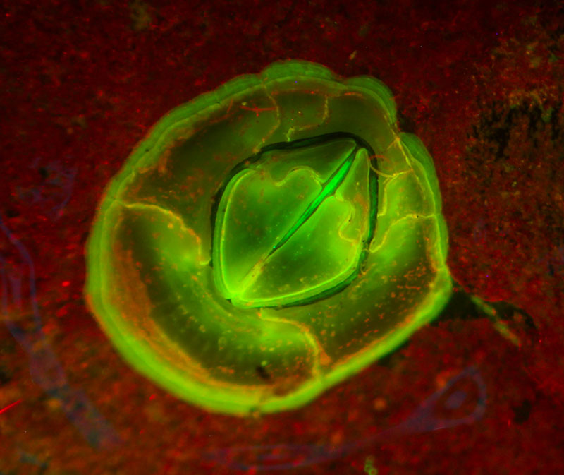
Coral
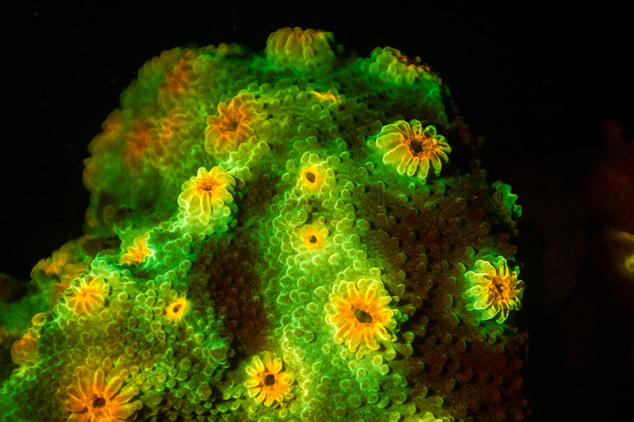
Coral recruits on settlement tiles, under the microscope
Fluorescence is a powerful tool in the study of juvenile corals and coral recruitment. Specimens that are hard to see in white light jump out in fluorescence.

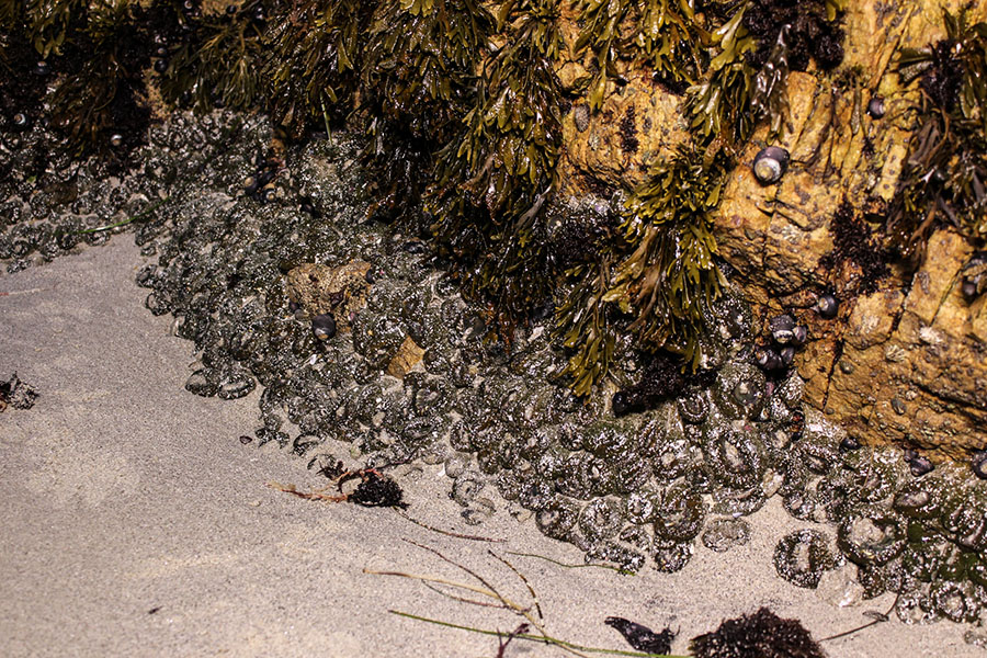
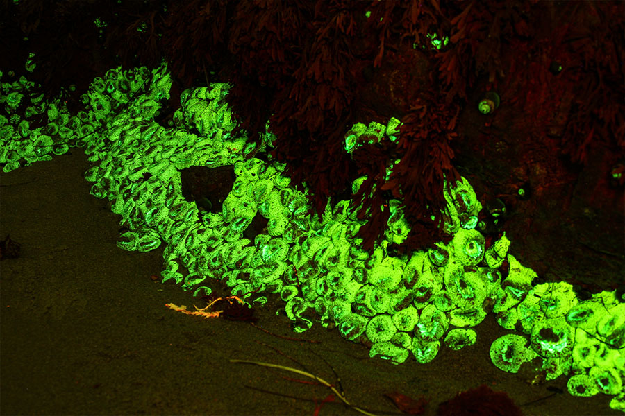
Barnacle
Industry
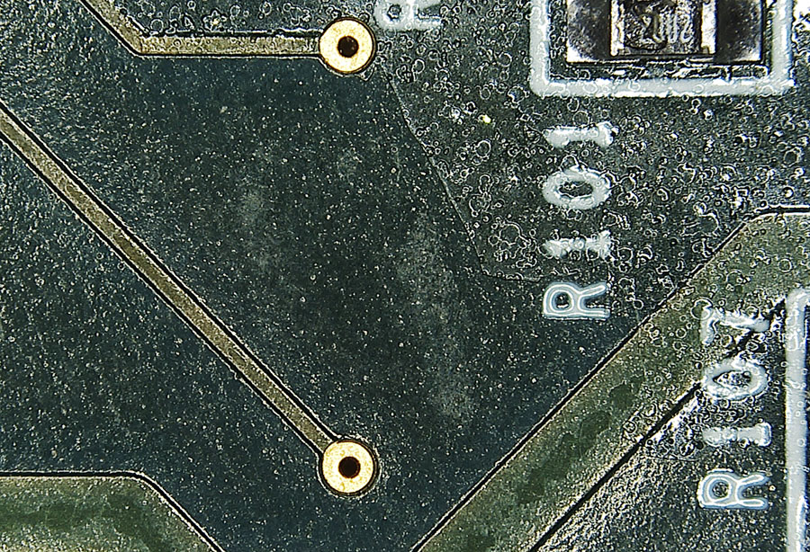
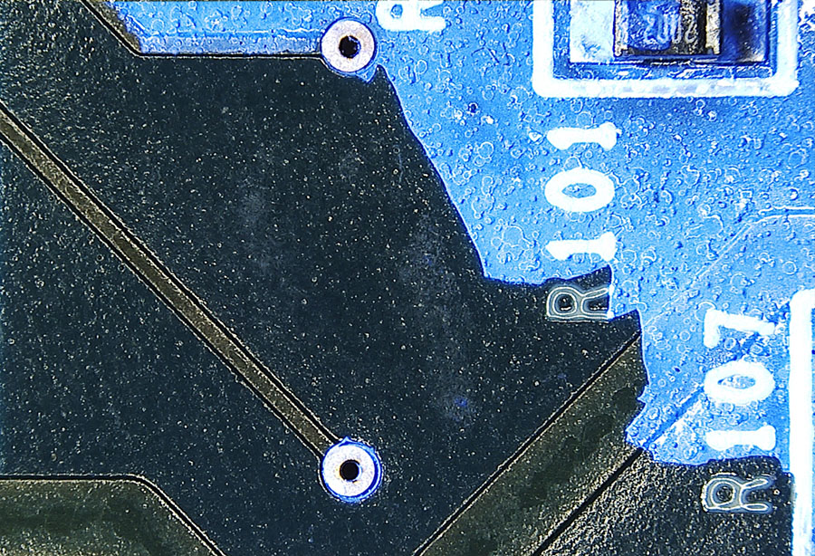
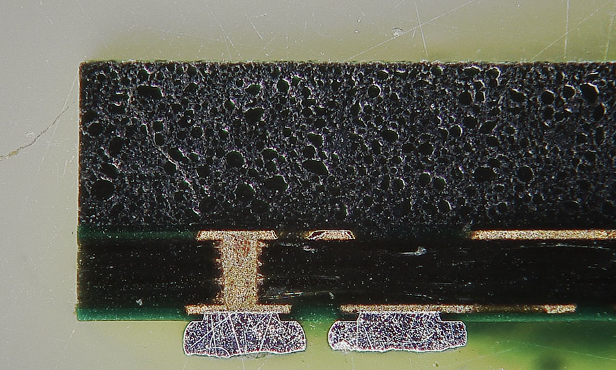
Electronics – Conformal coating on a circuit board
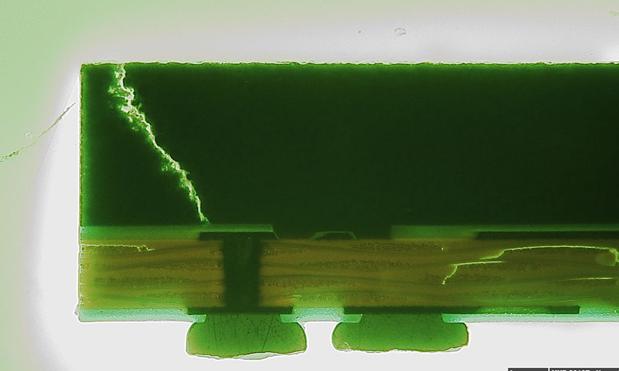
Failure analysis – Cracks in a cross section of an integrated circuit chip
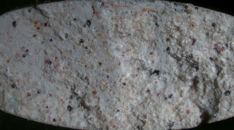
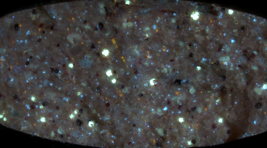
Pharmaceutical – cross-section of a multivitamin pill
Geology
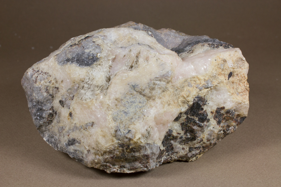
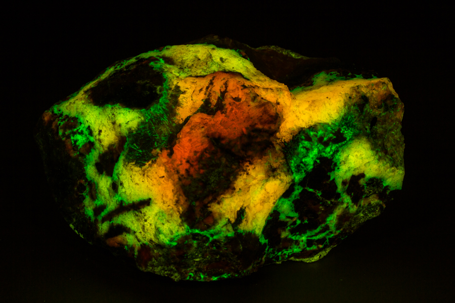
Mineral fluorescence – Calcite and Willemite, blue light excitation
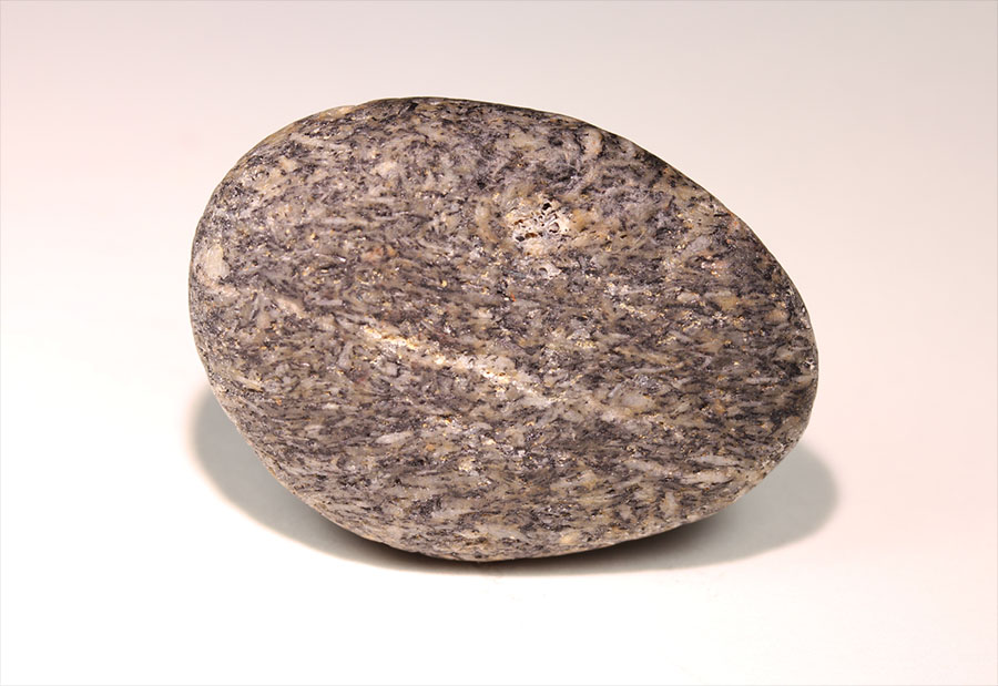
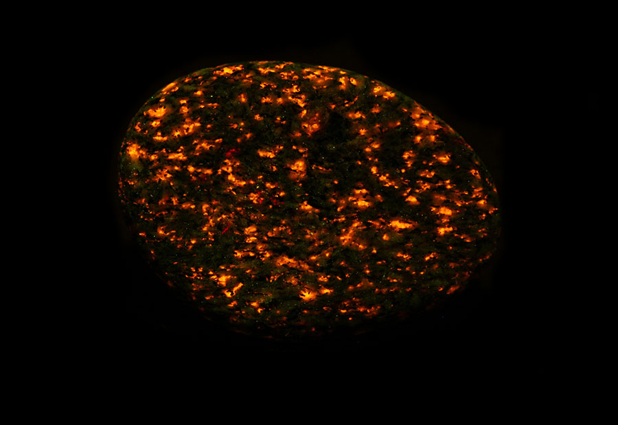
Mineral fluorescence – Sodalite
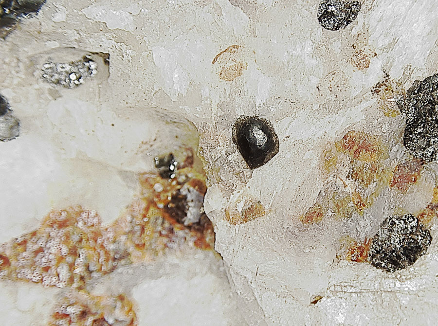
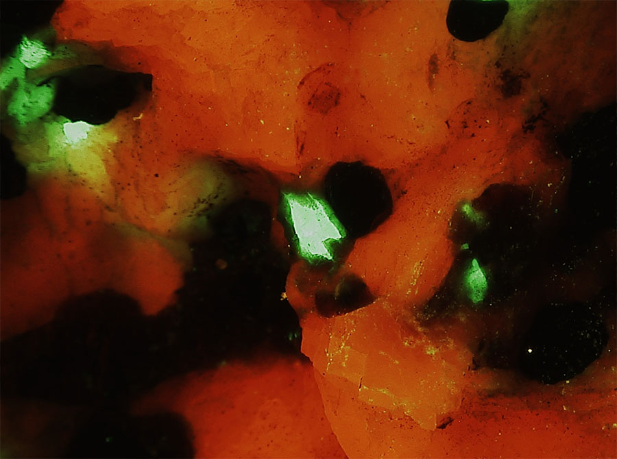
Mineral fluorescence – Specimen with calcite (red fluorescence) and willemite (green fluorescence) under the microscope
Mineral fluorescence – Gypsum – Video showing white light appearance, plus fluorescence under shortwave UV, longwave UV, and blue light
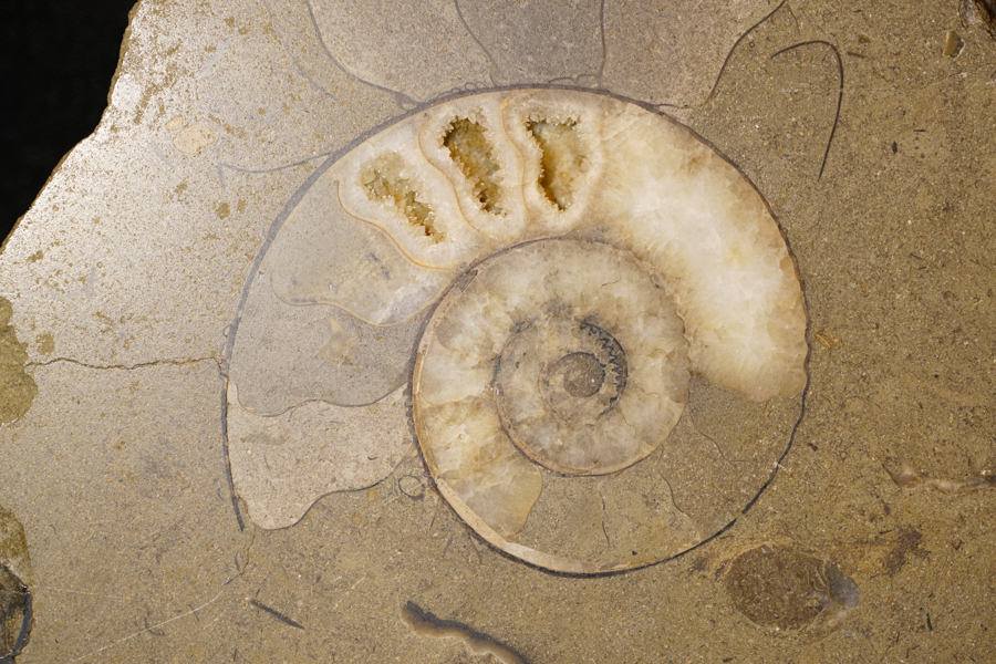
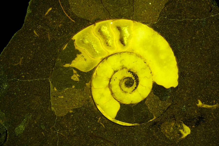
Ammonite fossil – calcite accumulation
Entomology
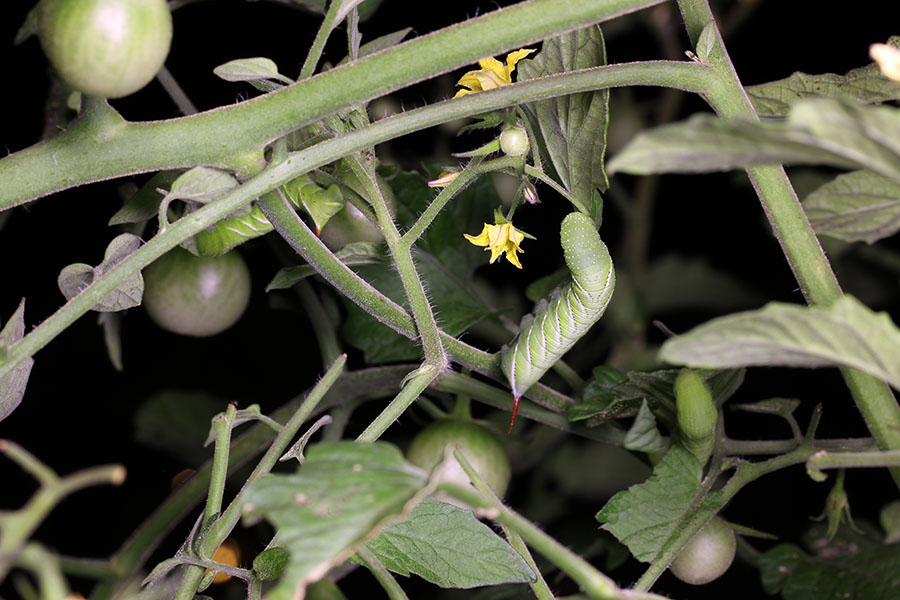
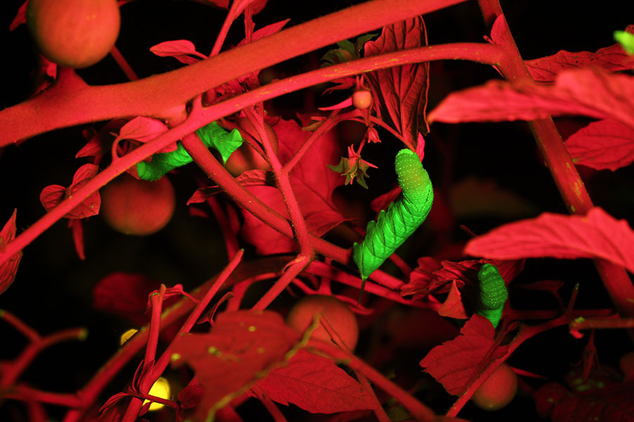
Tobacco hornworms on tomato plants
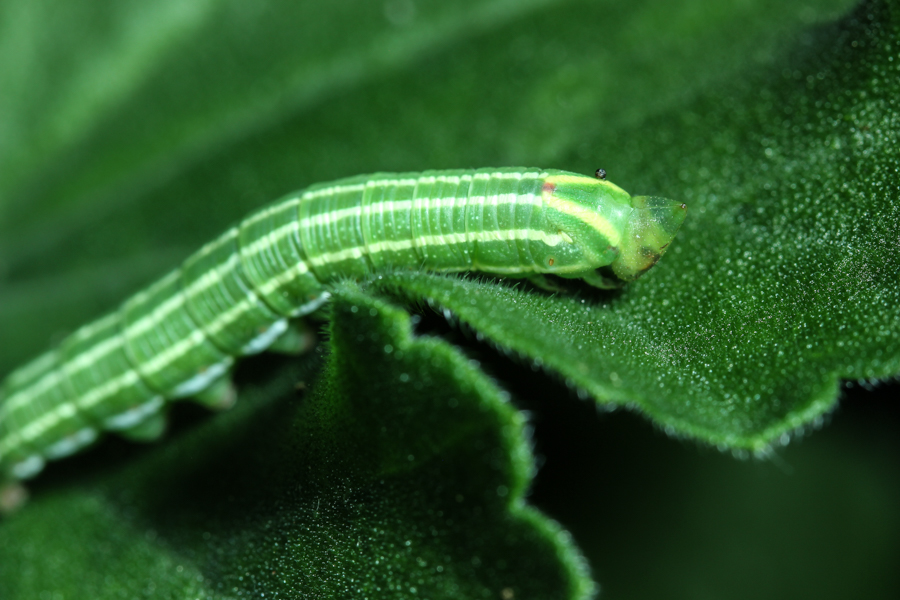
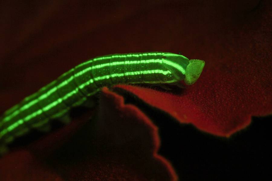
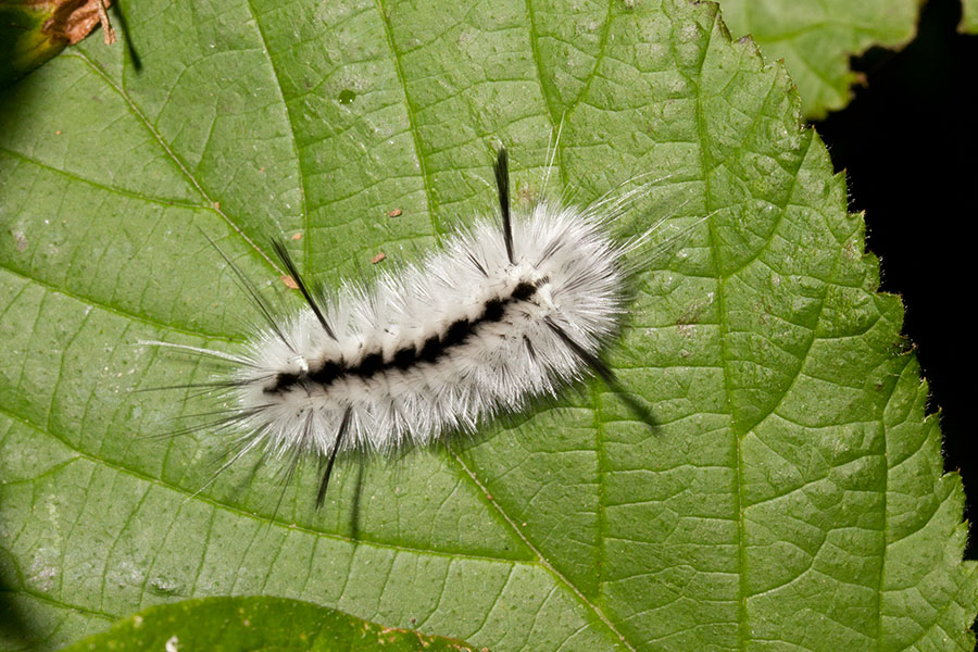
Caterpillars on leaves
Exploration
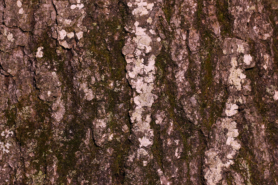
Tree bark
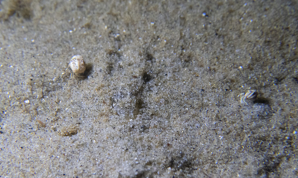
Low tide, Asilomar State Beach, California
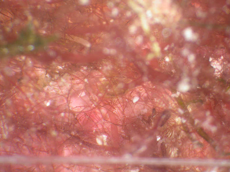
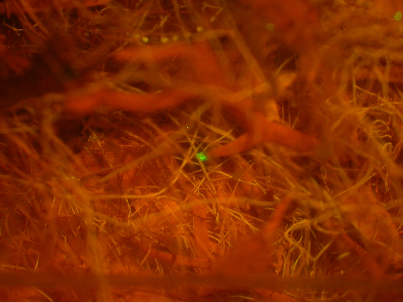
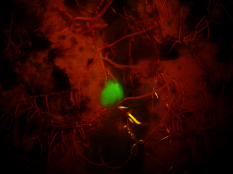
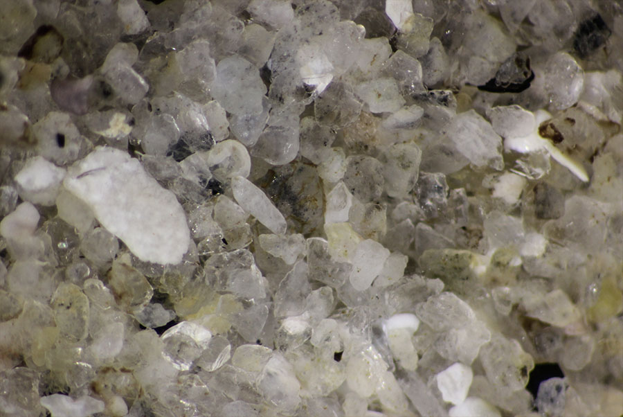
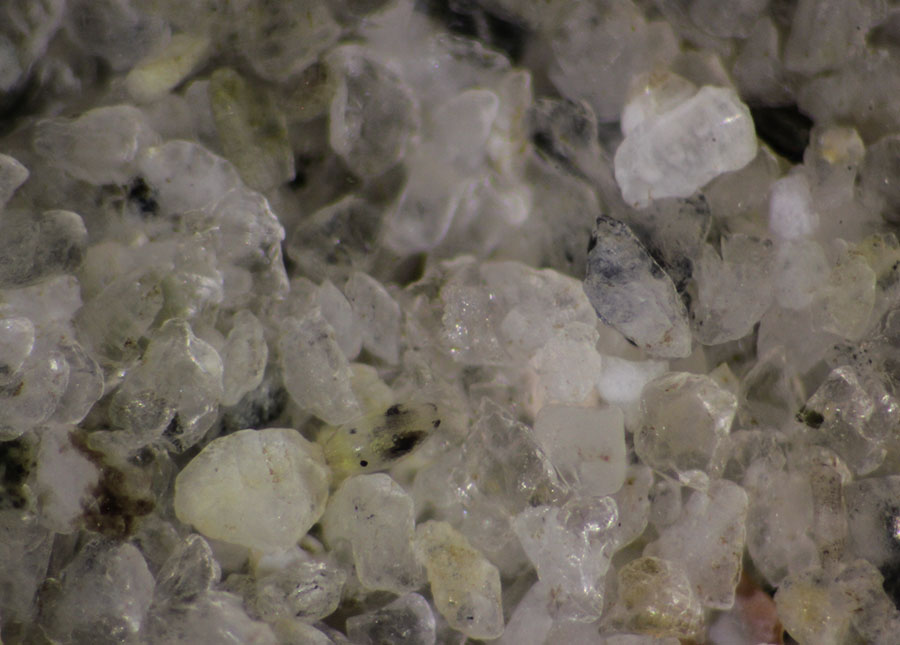
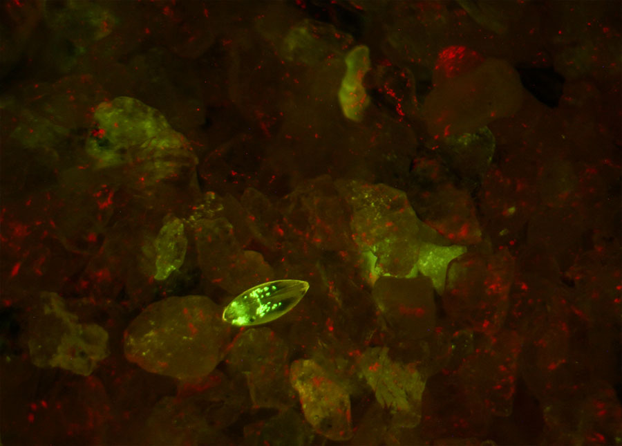
Sand scoop from Asilomar State Beach tide pool under the microscope
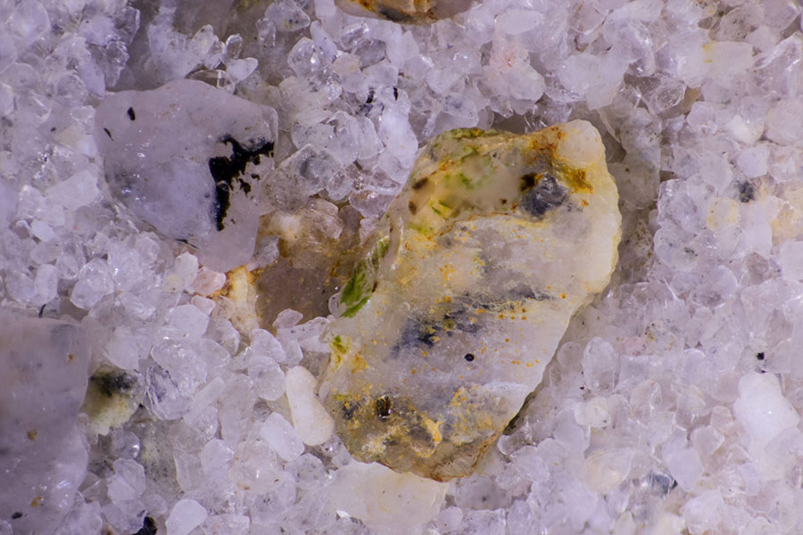
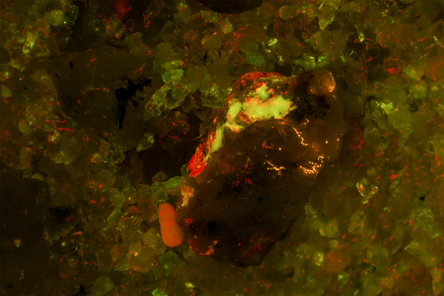
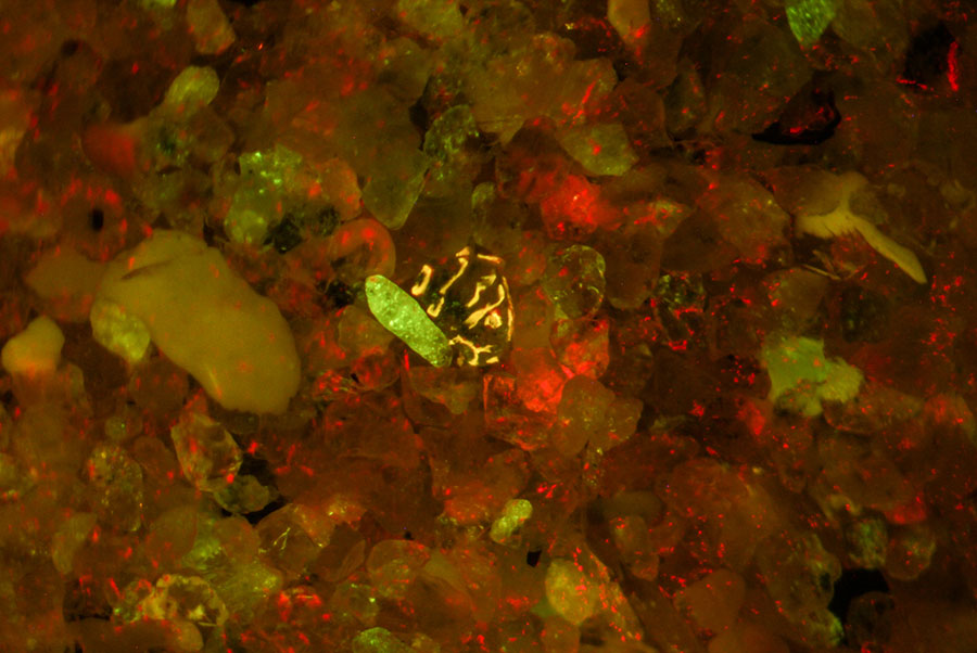
Find the ostracod!
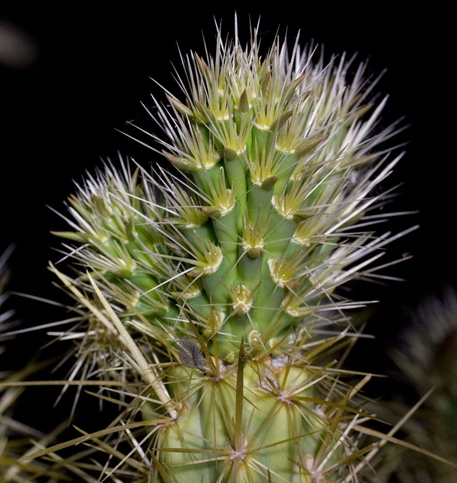
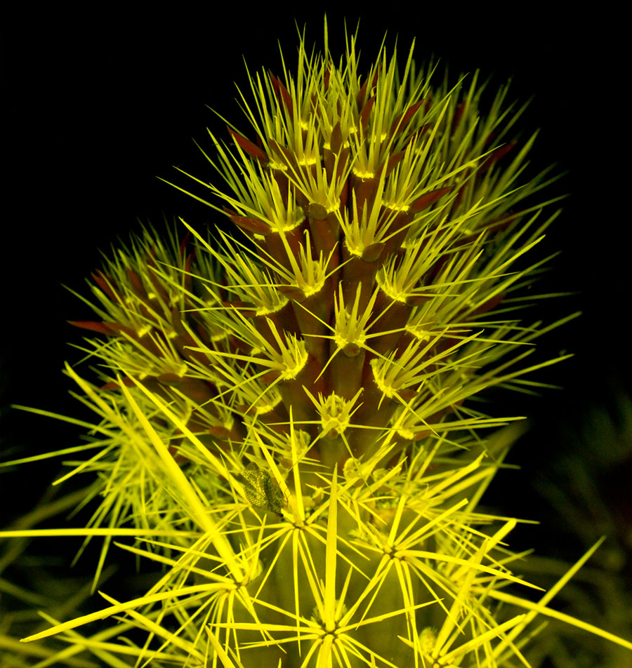
Cactus, Anza Borrego State Park
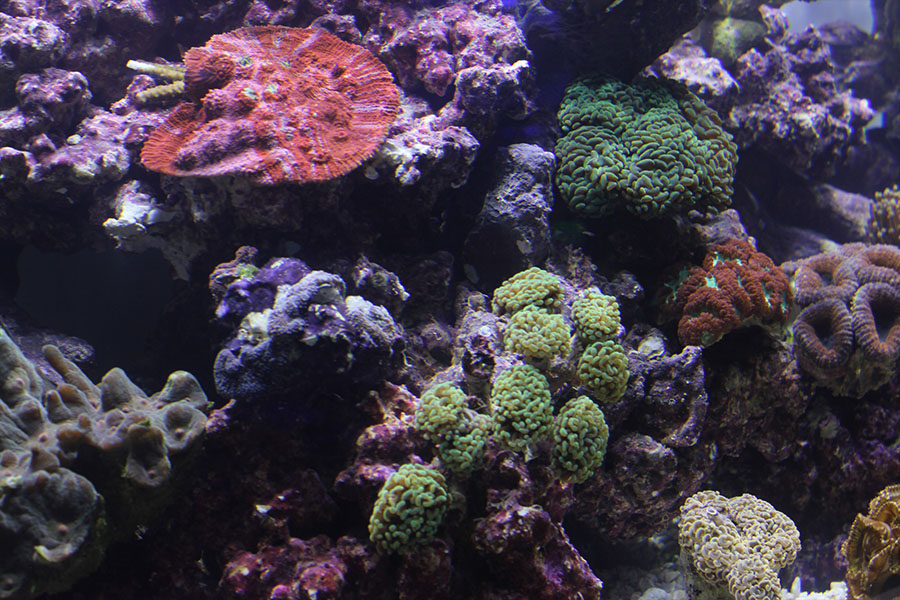
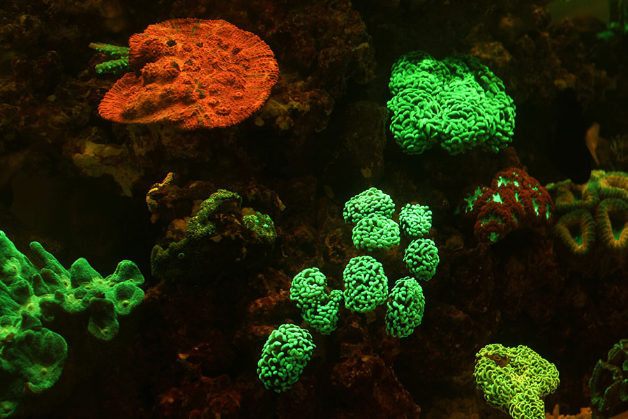
Aquarium
Reef aquarium