
Fluorescence of Cleared and Stained Specimens
We received this beautifully detailed fluorescence image of a seahorse skeleton from Kevin Conway at Texas A&M University. The specimen, a dwarf seahorse (Hippocampus zosterae), was prepared using a technique called ‘clearing and staining’, a method that produces an intact specimen with cartilage stained blue and bones stained red. It is a great way visualize the entire structure of an organism. The specific technique used was the one outlined in Smith et al., 2018 – see the links at the end of this article.
(Click image for larger view)
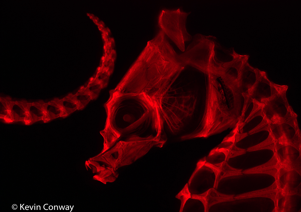
Cleared and stained seahorse – (c) Kevin Conway
The stain used for the skeleton is Alizarin Red S. It can be viewed under white light, but under appropriate excitation it glows a beautiful red. The image was made on a stereo microscope outfitted with the NIGHTSEA Stereo Microscope Fluorescence Adapter, using the Green (GR) excitation source and matching red barrier filter.
Kevin is an Associate Professor and Curator of Fishes in the Department of Ecology and Conservation Biology. His own research on comparative anatomy and development is focused on freshwater fish, and he just took this picture for fun to practice with the fluorescence setup. Thanks for sharing, Kevin!
Relevant links:
- Open access – Smith, W. L., Buck, C. A., Ornay, G. S., Davis, M. P., Martin, R. P., Gibson, S. Z., & Girard, M. G. (2018). Improving Vertebrate Skeleton Images: Fluorescence and the Non-Permanent Mounting of Cleared-and-Stained Specimens. Copeia, 106(3), 427-435.
- Open access – Girard, M. G., Davis, M. P., & Smith, W. L. (2020). The Phylogeny of Carangiform Fishes: Morphological and Genomic Investigations of a New Fish Clade. Copeia, 108(2), 265-298.
- Blog post (Southern Fried Science) on clearing and staining
- Web page at the American Museum of Natural History about the technique