
Python Skin Fluorescence
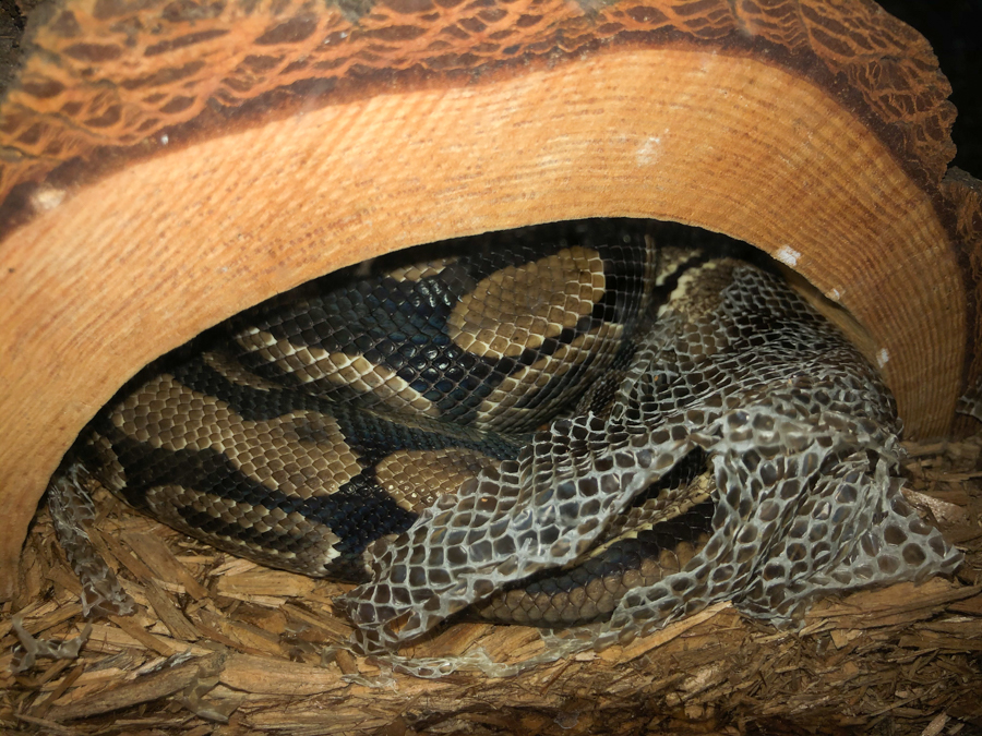
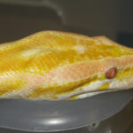
Here at NIGHTSEA we love new opportunities to explore fluorescence in the world around us. Recently, one of our staff was asked to snake-sit for her neighbors and had the pleasure of hosting Nagini – a 10 year old ball python (Python regius). Heather noticed that Nagini shed her skin shortly after arrival. Adult ball pythons only shed 2-4 times per year, so this was quite a chance event. Of course, Heather brought in some of the shed skin to so that we could ask our usual question – “Does it fluoresce?”.
(Click any image for larger view)
-

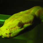
- Nagini showing her owner Spencer some love

-
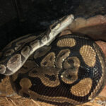
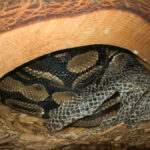
- Nagini at home

-
- Shed python skin
We illuminated the skin with longwave ultraviolet (UV) light and saw a striking blueish white fluorescence, as you can see in the image below. This image is reminiscent of the kind of picture we have seen for fluorescing scorpions, platypuses, flying squirrels, Tasmanian devils, and more. It is tempting to speculate that this fluorescence might have some importance in the visual life of the snake. Does it help it find a mate? Is it a visible warning to predators? Does it enhance camouflage in some lighting conditions? These are the kinds of questions that we commonly see asked with new observations of fluorescence. [Side note – it is not uncommon for these articles to say that the animals in question ‘glow in the dark’. They don’t 🙂 See our article on that here.]

Shed python skin fluorescing under UV light
We expect, though, that a visual function is pretty unlikely. Snake skin is largely made of the protein keratin, the same material as our own hair and nails. For a quick visual comparison, we added a standard issue human hand into the scene (don’t worry, the rest of the human was still attached, and no one was harmed in the making of this picture) and found that the fluorescence of the fingernails was pretty much equivalent to that of the snake skin. Although this makes for fun photographs, it is hard to imagine that the fluorescence of our fingernails serves a visual function – rather it is an incidental, and minor, byproduct of the material used to create them.
-
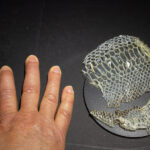
- Hand and python skin, white light

-
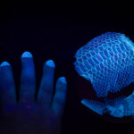
- Hand and python skin, UV light

As another example, see the picture below that includes the snake skin, fingernails, and … teeth. Our teeth are not made of keratin, but like fingernails it is hard to imagine that this fluorescence, while quite striking and great for taking a fun photo, is functional.

The fluorescence from NIGHTSEA employee Cat’s pearly whites under UV light is much brighter than that emitted by python skin or fingernails.
Technical interlude – Spectral exploration
[Feel free to skip this section to move on to more discussion below as to whether the fluorescence might have a visual function]
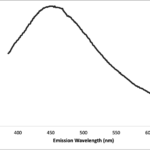
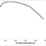
We did a deeper dive into the spectral characteristics of the fluorescence using our old FluoroMax-2 spectrofluorometer. The graph below shows the emission spectrum of the python skin when excited with a wavelength of 360nm (about the same as the flashlight used for the pictures). It shows a fairly broad emission with a peak at about 450nm, in the blue. A measure of the excitation spectrum (right) shows a broad curve, peaked in the 370 – 400nm region. This kind of data helps you understand what light is going into the fluorescing substance, and what is coming out.
-
- Emission spectrum, excitation at 360nm

-
- Excitation spectrum, emission at 500nm

We also tried exciting the python skin at a different wavelength to see what happens. The graph below shows the emission spectrum for excitation at 450nm, added to the prior graph. We can see that the peak emission is now shifted to a new, longer wavelength.

Emission spectra, excitation at 360 and 450nm
This shift in the emission peak suggests that whatever is fluorescing in the python skin has more than one underlying component. The physics behind fluorescence (see, for example, the article on this web site about the Jablonski diagram) tells us that for a given fluorescing molecule the emission spectrum will be independent of the excitation wavelength. So when we see the kind of shift above we know that more than one component must be present.
We measured a series of emission spectra at excitation wavelengths ranging from 340 to 650nm, at 10nm intervals, to create what is called an excitation/emission matrix, and then plotted the data in a contour plot (below). The emission wavelength is on the horizontal axis, the excitation wavelength on the vertical axis, and the color shading indicates relative intensity. We added the sloping solid line connecting the emission wavelength peaks for each excitation wavelength. If there had been a single fluorescing substance this line would have been vertical, extending straight up from the wavelength of maximum emission on the horizontal axis.

Excitation/emission matrix. Blue line indicates peak emission wavelength for each excitation
What are these multiple substances contributing to this complex fluorescence? We don’t really know. Besides keratin, we know that there must be other components present, like pigments that give Nagini her colors and spots. This bears further investigation, beyond our scope here at NIGHTSEA.
Might the fluorescence have a visual function?
Let’s come back to the question of a visual function. If so many creatures and compounds fluoresce when exposed to ultraviolet or other light, how might we determine when biological fluorescence is functional rather than merely incidental?
Justin Marshall and Sonke Johnson, in their excellent paper ‘Fluorescence as a means of colour signal enhancement’ [Marshall, J., & Johnsen, S. (2017). Fluorescence as a means of colour signal enhancement. Philosophical Transactions of the Royal Society B: Biological Sciences, 372(1724).], lay out a checklist of things to consider in regard to the potential for fluorescence to have an ecological significance:
- Is there a fluorescent compound (fluorophore) present in a visible location?
- That is, does the fluorescence occur in a part of the body that is visible either generally, or during a specific display behavior?
- What are the excitation and emission wavelength ranges of the compound/tissue?
- This data is necessary to understand how ambient light in the natural environment can stimulate the fluorescence, and for quantitative modeling.
- What are the spectral sensitivity ranges of potential viewers?
- Something has to be able to see the fluorescence in order for it to be relevant!
- Under what natural lighting conditions is the fluorescence viewed?
- This affects both how strongly the fluorescence might be excited, and how much contrast there might be against the background.
- Are there visual behaviors that might either rely on or be assisted by the fluorescence color or pattern?
- Even if fluorescence is in a visible location, is strong, and can be seen by a viewer, you still have to demonstrate that the fluorescence is being used.
The same factors are presented in a slightly different way in a paper by NIGHTSEA founder Charles Mazel [Mazel, C. (2017). Method for determining the contribution of fluorescence to an optical signature, with implications for postulating a visual function. Frontiers in Marine Science, 4, 266.]. Both of these papers show the mathematics involved in computing the fluorescence emitted by the subject under different lighting conditions.
Let’s look at the criteria above in the context of Nagini’s shed:
- Yes, fluorescence is present and it seems to be throughout the skin.
- The spectral data we have presented here shows what light we expect to excite the fluorescence, and what light will be leaving the surface. This in itself is not enough to compute the expected contribution of fluorescence to the signal under arbitrary lighting conditions – we would also need to measure/compute the efficiency of conversion of incident energy to fluorescence.
- At this time we do not know what the ‘potential viewers’ are. Other ball pythons? Other animals that they might interact with? Pit snakes such as pythons have notoriously poor vision. As nocturnal animals, they instead rely on two special organs to sense the world around them. The first is the Jacobson’s organ, located on the roof of their mouth that allows them to “smell” chemical signals from prey and other environmental stimuli. The second is unique to pit snakes and is known as the pit organ. Located in a hole in their head, a pit organ detects heat in the form of infrared radiation and allows the snake to “see” its predator or prey.
- Ball pythons are largely nocturnal, tending to hide during the day! This in itself argues against a visual function for the fluorescence.
- There are no obvious, or even likely candidate, functions that might be affected by fluorescence, but that would have to be determined by ecological observation and experiment.
One question in the Mazel version of the criteria list is ‘Is there enough energy transferred into the fluorescence band to make a meaningful contribution to the visual signal under relevant illumination conditions?’ In order for there to be fluorescence there has to be an illumination source. The fluorescing surface will reflect some of the incident light and will absorb some light and transfer it to new wavelengths by fluorescence. The light leaving the surface will be a combination of the reflected and fluoresced components, and only in exceptional circumstances is the fluorescence portion likely to be significant relative to the reflected portion. And if the fluorescence is really quite weak, as the photographs that include fingernails and teeth indicate, then it is much more likely that the fluorescence of Nagini’s skin is an interesting curiosity, and not an ecologically important property.
Another question that is always interesting to ask is, “Does the fluorescence reveal any patterns hidden from the naked eye?” It would make a visual case much more compelling if there were patterns that could only be viewed in the spectral range of the fluorescence. But as far as Nagini – and most observations that we have seen of fluorescence in various animals – is concerned, the short answer is no. If we look back at the UV-excited photograph of Nagini’s skin, the fluorescence is fairly uniform across her scales. In fact, this piece of skin was from one of her lighter patches (as seen in her head shot above). The darker area of skin did not fluoresce. So the dark light pattern that we see would not be changed by fluorescence.
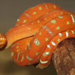
Here are some additional images of snake fluorescence, taken in a well-stocked Florida pet store. The fluorescence in this case was excited by blue light and photographed through a yellow filter, so the fluorescence of the skin does not have the same bluish-white appearance as when taken under ultraviolet light.
-
- white light

-
- fluorescence under blue light

-
- white light

-
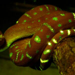
- fluorescence under blue light

Final Thoughts
Unfortunately we did not have an opportunity to observe and photograph the fluorescence of Nagini’s skin while she was still wearing it. Based on snake behaviors and spectrofluorometric data, it seems unlikely that Nagini’s “glowing skin” serves a biological function. What we hoped to do with this article was not to proclaim the next big discovery about the eyesight of pit vipers. Instead, we wanted to demonstrate a line of questioning and investigation that can be applied to research in the visual function of fluorescence. Not to mention, we wanted some fun pictures.
And a parting suggestion to those documenting new instances of fluorescence in various animals – include someone’s hand in the picture, or better yet, have someone join the photo shoot and smile. That will give you a better idea of what the intensity really is.
