Signal from Noise – A Cautionary Fluorescence Tale
Posted by NIGHTSEA founder Charles Mazel
Preface – a lesson from the past
A very long time ago I worked in deep sea survey, spending weeks at a time at sea on big ships mapping the bottom with sound. On one of my first trips one of the engineers from our group pointed to an oscilloscope trace from a hydrophone suspended below the ship. It showed signal intensity as a function of sound frequency. The trace looked something like the figure below.

This looked easy to interpret – there was mostly noise but at one particular narrow frequency range there was a distinct signal. But then he twiddled some dials and it went away – he was just fooling around and making a point. He had applied a narrow-band frequency filter to the detector output and then provided gain, amplifying the signal. The result was a distinct spike at the frequency setting of his filter and a weak noise signal outside it. But there was really nothing special going on at that frequency. It was just noise being very selectively amplified.
The lesson stuck with me, always in the back of my mind. Filters can be valuable, but when applied without thought and understanding they can create false readings, the apparent presence of signal when there is only noise. This has served me well throughout my career, especially since in a large part of that I was constantly using filters to view fluorescence. Whenever you do that you have to think not only about what you are seeing, but what you are NOT seeing because of the filter. I have seen mistakes, even in the peer-reviewed scientific literature, where the ‘results’ do not tell the full story due to a lack of this awareness.
The present – a spectral mystery solved
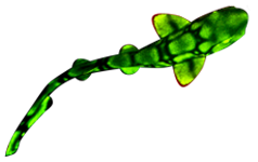
In early 2021 I was in San Diego, CA, exhibiting our Stereo Microscope Fluorescence Adapter at the annual Drosophila Research Conference. My good friend Joseph Daniels from Archon Scientific, purveyors of fine fly food, brought a researcher to my display booth. She was using both green and red fluorescent proteins in her research, with the aim of visualizing features in her flies. Her images were being ruined by a background fluorescence from something in the fly guts. There was a strong green fluorescence when looking at Green Fluorescent Protein (GFP), and a red fluorescence when looking at a red fluorescent protein (I forget exactly which one). She suspected that it might be fluorescence from the fly food and had gone to Archon to ask them about that. Why would the fly food fluoresce red and green? They did not know, and brought her over to me.
Drawing on that lesson from the past I only needed to ask a few key questions to get to the root of the problem:
How was she doing the imaging?
She was using confocal microscopy. This is a very sensitive high magnification technique using lasers for fluorescence excitation and sensitive detectors for signal capture. A great way to capture even weak fluorescence signals.
What kind of filters does the microscope have – longpass or bandpass?
The answer, as expected was bandpass. High-end microscopes like a confocal typically use narrow-band detection filters so that they can capture the fluorescence they are looking for while rejecting background. That usually works well, but not in this case.
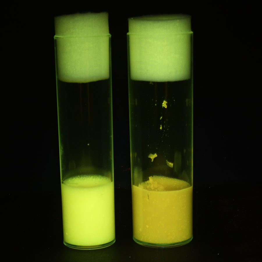
I borrowed a couple of vials of fly food from Joseph’s booth. I had filter sets for looking at green and red fluorescence with me and guess what – the Royal Blue excitation in combination with a longpass barrier filter elicited a distinct yellow fluorescence, while the Green excitation with red barrier filter resulted in a nice red fluorescence. With the Royal Blue, if we had been looking through a bandpass filter we would have seen only green.
Mystery easily solved. The fly food has a very broadband fluorescence, but when viewed through a narrow filter with strong excitation and a sensitive detector it looks the same as a ‘true’ fluorescence of that color. Signal created from noise.
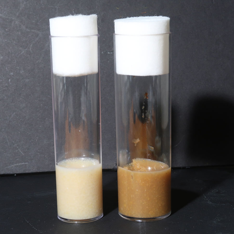
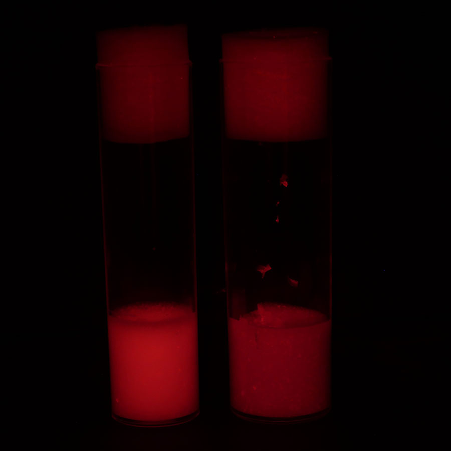
I brought vials of two different kinds of fly food back to my office and photographed them (below) with white light and then with Ultraviolet, Royal Blue, and Green excitations, viewing through a longpass barrier filter in each case. Nice fluorescence, with the apparent color dependent on the filter being used.
-

- Fly food, white light
-
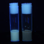
- Fly food, Ultraviolet excitation

-

- Fly food, Royal Blue excitation
-
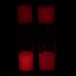
- Fly food, Green excitation
This is the key take-away: If you are viewing fluorescence through a bandpass filter of any given wavelength and you see a fluorescence signal you have to keep in mind that there are two main possibilities:
- The source of the fluorescence is fluorescing primarily in that wavelength range and is the fluorophore that you want to see (GFP, YFP, RFP, etc.).
- The source of the fluorescence is fluorescing over a broad wavelength range but your filter only allows you to see the fraction of that emission that falls within the filter’s transmission band.