Conservation and Fluorescence
Fluorescence has diverse applications in the field of conservation, primarily to aid in understanding more about the subject that is being worked on, but also for other applications such as cleaning. The technique can be used with paintings, paper, objects, and more. We at NIGHTSEA are just beginning to learn where our fluorescence-enabling equipment can contribute to this field, at scales from macro to micro . There are a number of institutions that are already using our systems, and some have graciously shared application examples here.
NIGHTSEA equipment
Our primary strength is in supplying turnkey systems that add a fluorescence capability to existing microscopes, including:
- Stereo microscopes of virtually any make, model, and vintage
- Digital microscopes, including:
At the macro scale, we offer:
- Xite fluorescence flashlight system
- Solutions for photography (on request)
In addition to ultraviolet light (UV, 360-380nm), which is the most commonly used in the field, we offer excitation sources in 4 wavelength ranges: Violet (400 – 415nm), Royal Blue (440 – 460nm), Cyan (490 – 515nm), and Green (510 – 540nm). Ultraviolet is not always the best excitation source for any given fluorescing subject, and this variety provides opportunity for research, exploration and discovery.
Application examples
The images below include some that were provided by users of our system, and some that we made ourselves.
- Paint and lacquer cross sections
- Paintings and prints
- Touch-ups on a classic comic book cover
- Repairs
- Paper
There are many other potential applications of fluorescence in the field of conservation and we will add to this page as we acquire additional examples.
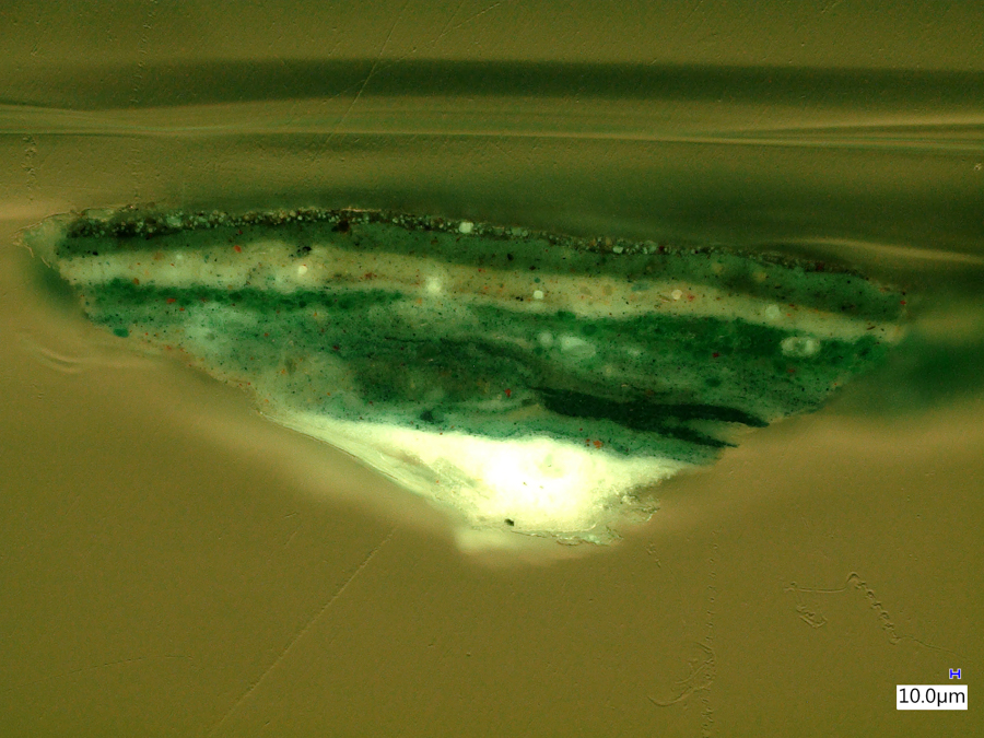
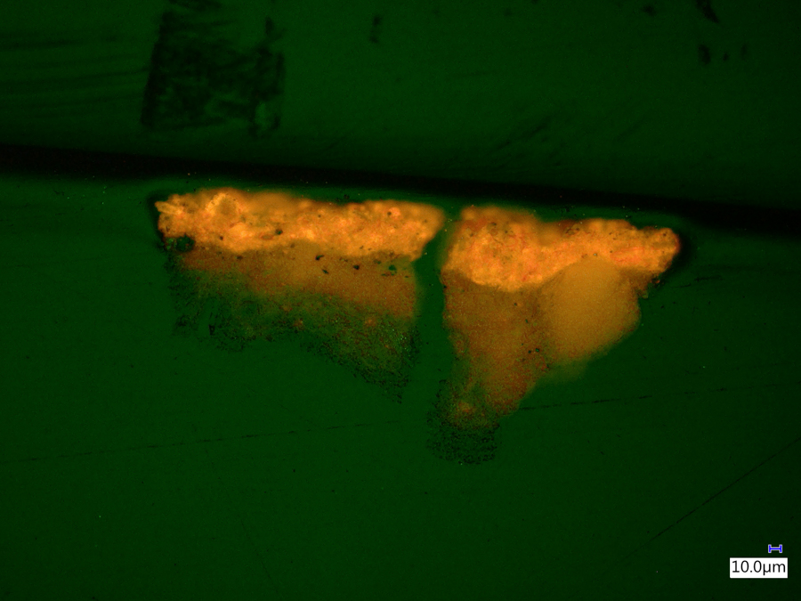
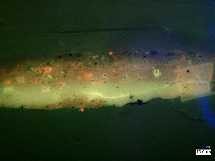
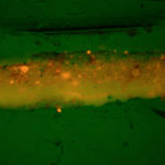
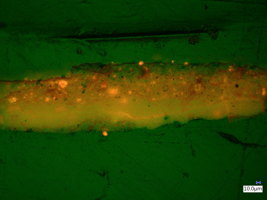
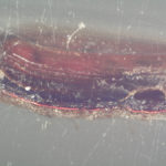
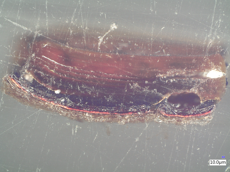
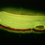
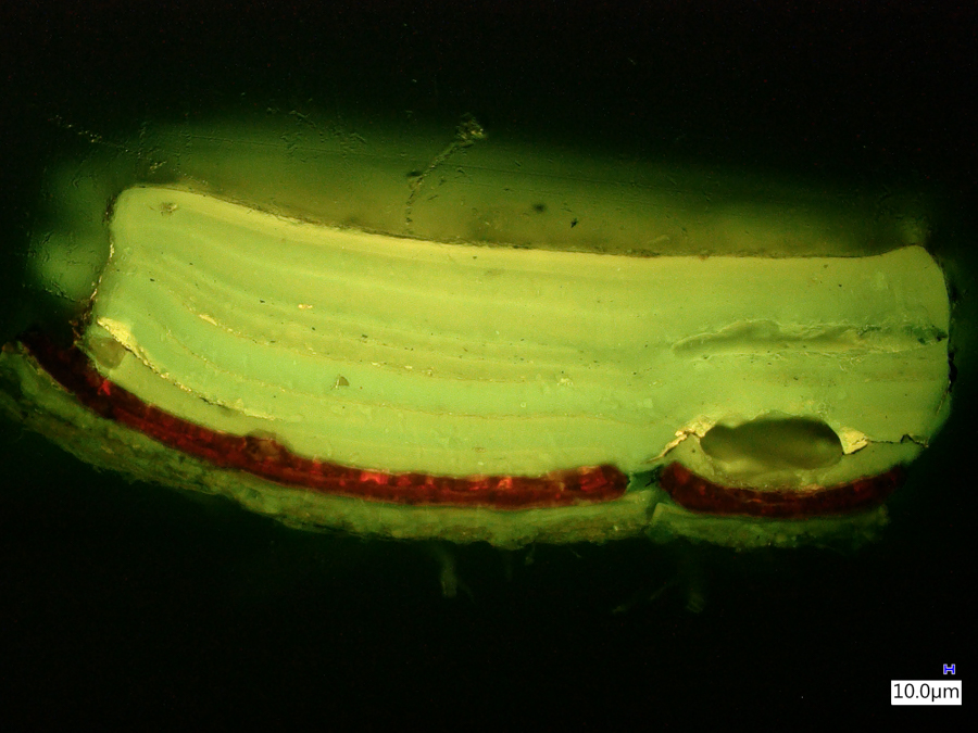
Paint and lacquer cross sections
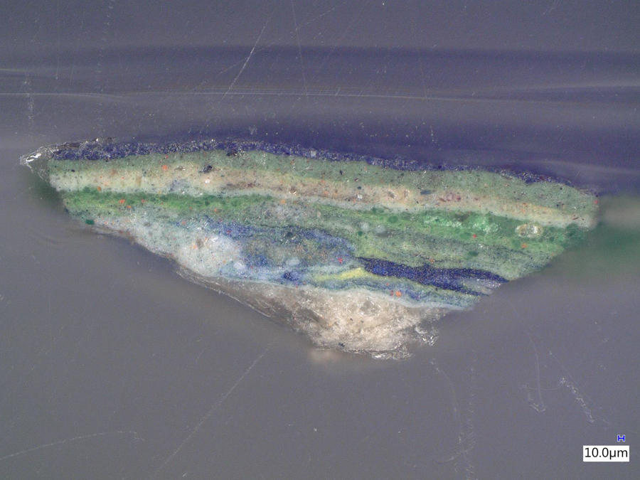
The images below were made at the Straus Conservation Laboratory at the Harvard Art Museum. They were made with the NIGHTSEA Royal Blue or Cyan excitation/emission sets, as noted, on a Keyence VHX-6000 microscope with the VH-ZST lens.
All images © President and Fellows of Harvard College
(Click any image for larger view)
-
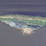
- Paint chip cross section, white light. #1951.49.
-
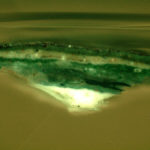
- Paint chip cross section, fluorescence, Royal Blue. #1951.49.
-
- Paint chip cross section, white light. #1939.111.
-
- Paint chip cross section, fluorescence, Cyan. #1939.111.
-
- Paint chip cross section, white light. #1923.60.

-
- Paint chip cross section, fluorescence, Royal Blue. #1923.60.
-
- Paint chip cross section, fluorescence, Cyan. #1923.60.
-
- Lacquer cross section, white light. #1984.463.
-
- Lacquer cross section, fluorescence, Royal Blue. #1984.463.
Samples:
- Accession #1951.49. Poèmes Barbares, 1896, Paul Gauguin. Oil on canvas. Harvard Art Museums/Fogg Museum, Bequest from the Collection of Maurice Wertheim, Class of 1906.
- Accession #1939.111. Mummy Portrait of a Woman, c. 200 CE. Tempera on hardwood. Harvard Art Museums/Arthur M. Sackler Museum, Gift of Mrs. John D. Rockefeller, Jr.
- Accession #1923.60. Mummy Portrait of a Woman with Earrings, c. 130-140 CE. Encaustic on linden wood. Harvard Art Museums/Arthur M. Sackler Museum, Gift of Dr. Denman W. Ross.
- Accession #1984.463. Folio from an Illustrated Manuscript of a Compendium of Knowledge (Jung), made for Shah Sulayman, c. 1669-1670. Ink, opaque watercolor and gold on paper. Harvard Art Museums/Arthur M. Sackler Museum, Gift of Philip Hofer.
Paintings and prints
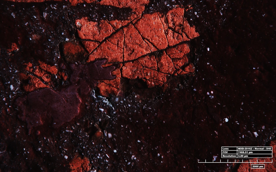
Identification of original 17th century paint vs. modern overpaint
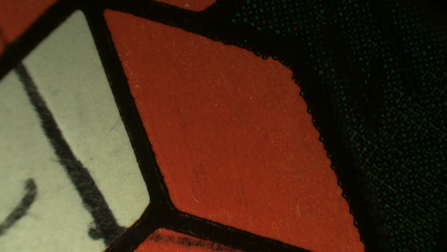
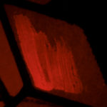
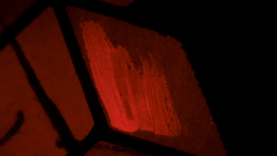
The two images below were acquired at the Eskenazi Museum of Art at Indiana University, Center for Conservation, using the Hirox RH-2000 Digital Microscope System and the NIGHTSEA Ultraviolet excitation/emission set. They were able to document an area of the painting ‘Joseph Seeking His Brothers‘ (Eustache Le Sueur, ca. 1647–1650) with some remaining original 17th century red oil paint (likely vermilion) surrounded by modern (approx. 50 years old) overpaint. The original paint exhibits a red fluorescence (below right), while the overpaint does not (white light image, below left).
Images © Eskenazi Museum of Art at Indiana University, Center for Conservation
-
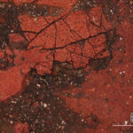
- Painting detail, white light

-
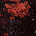
- Painting detail, fluorescence
————
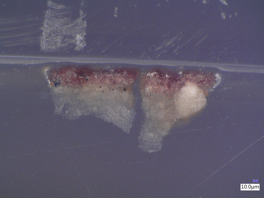
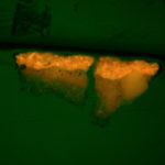
Analysis of an area of paint loss
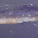
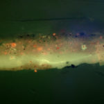
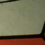
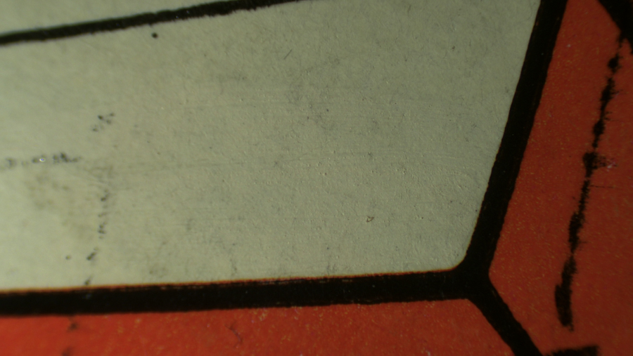
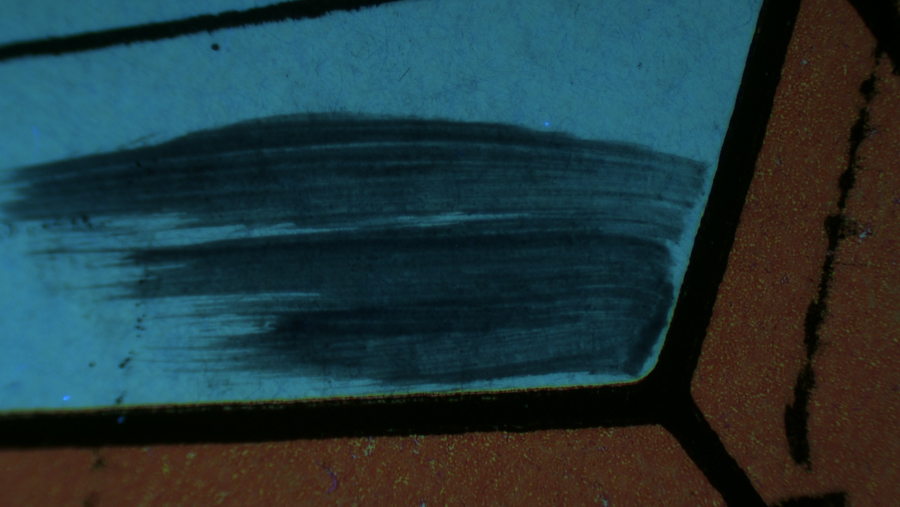
The images below were acquired at the Heritage Conservation Centre, National Heritage Board, Singapore, using a Keyence VHX-6000 microscope with the VH-Z20 lens mounted on a mobile stand and a NIGHTSEA UV excitation/emission set. Thin brown varnish over zinc white ground. The fluorescence of the patch that appears fairly uniformly pale in the white-light image (left) arises from zinc white. The difference in brightness of the UV-excited fluorescence results from a later retouching/alteration. It was found that the more brightly fluorescent area (1) was older than the less fluorescent area (2).
Images © National Heritage Board, Singapore
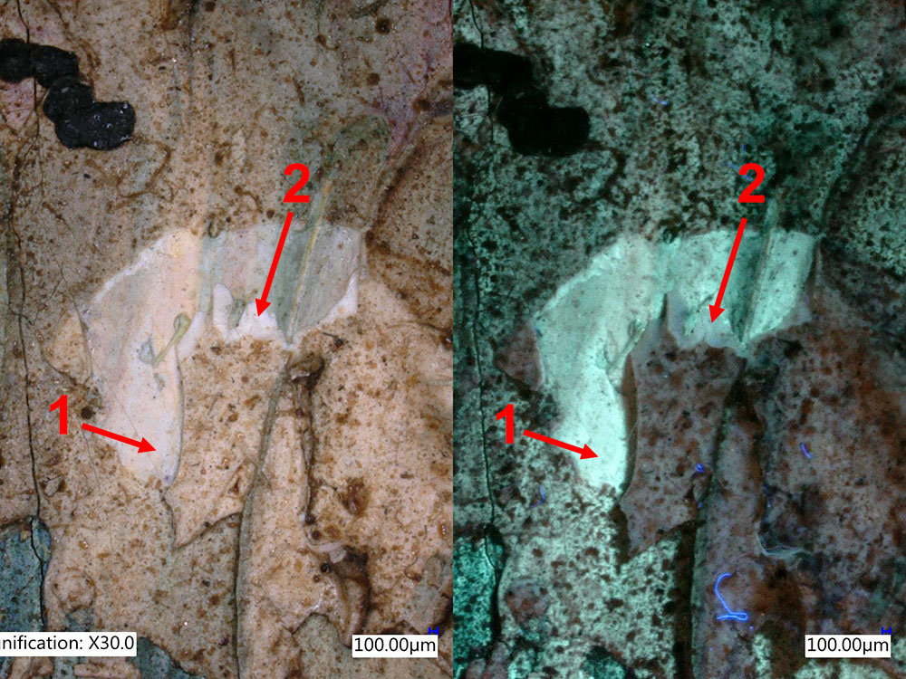
Paint touch-up, white and UV light.
Source:
- “Bunga Crisant 1 (Chrysanthemum)”, by Emiria Sunassa, c.1946-1949. Oil on hardboard
————
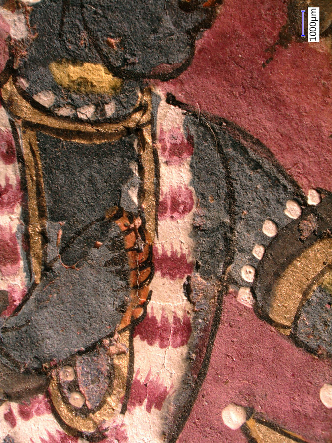
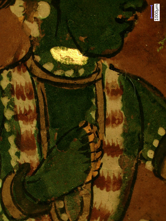
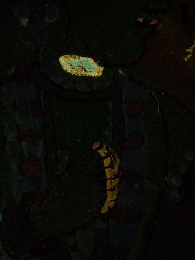
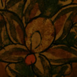
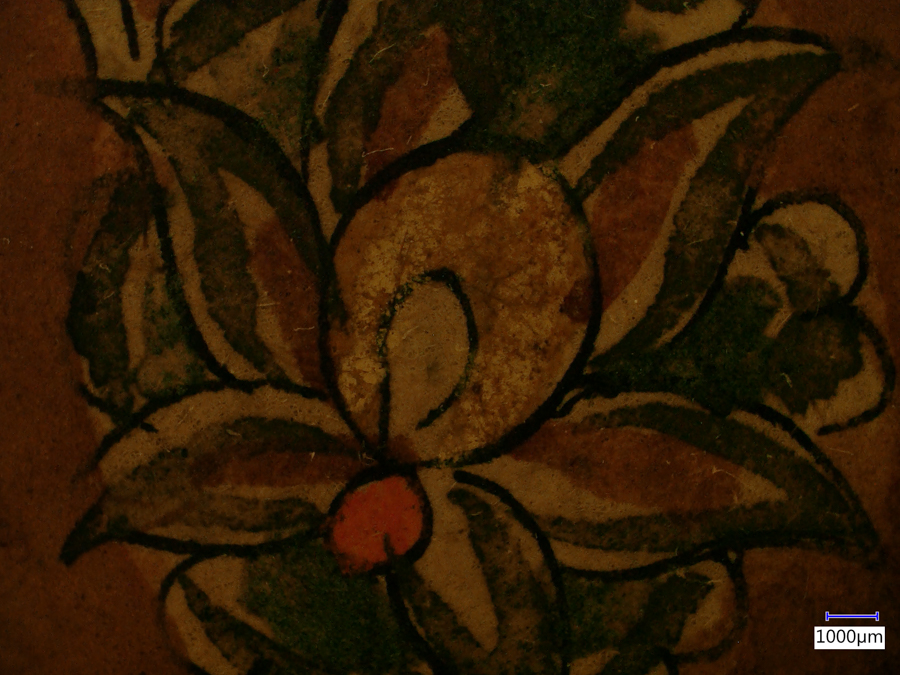
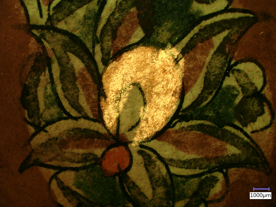
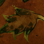
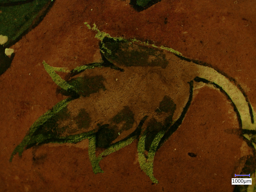
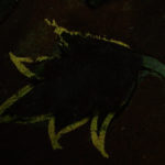
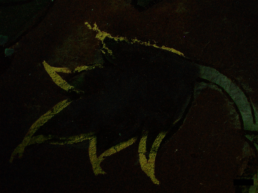
Fluorescent pigments in a 17th century Indian painting
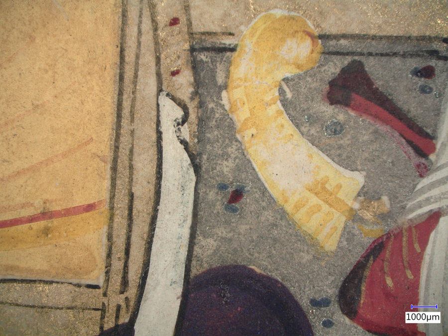
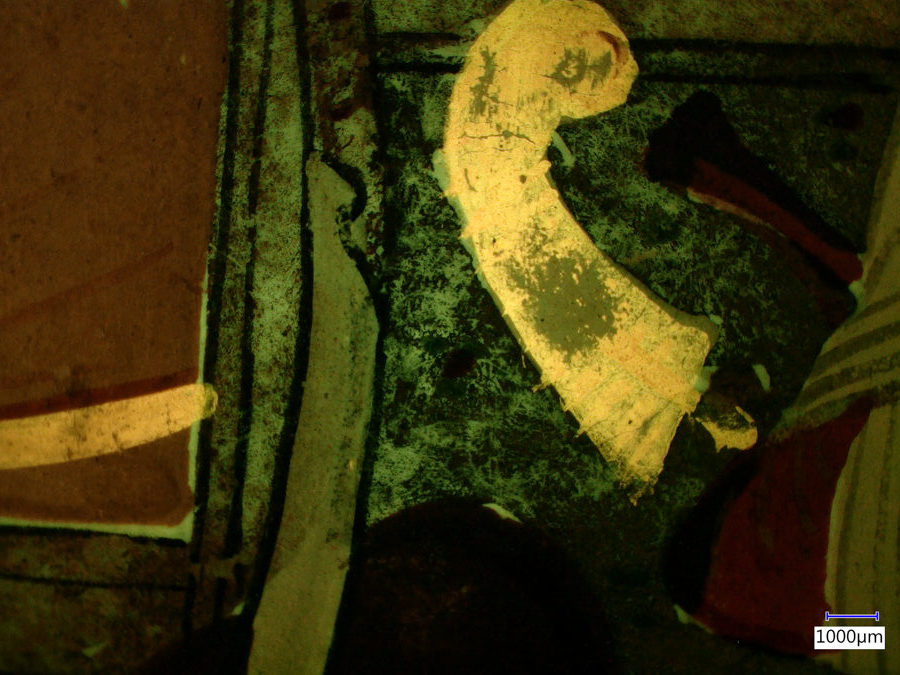
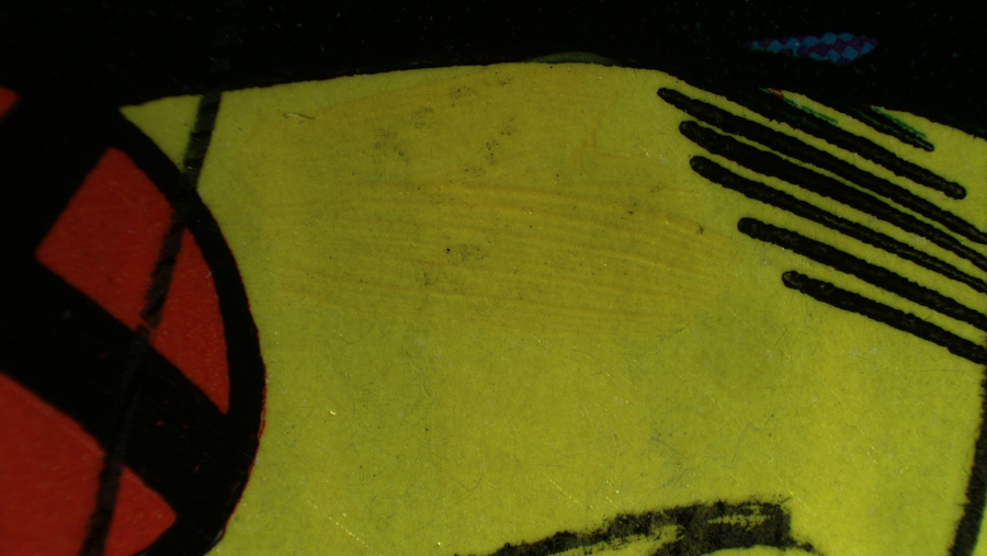
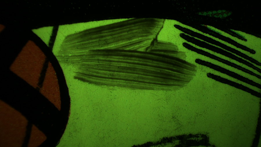
The images below were made at the Straus Conservation Laboratory at the Harvard Art Museum. They were made with a Keyence VHX-6000 microscope with the VH-ZST lens and the NIGHTSEA Royal Blue excitation/emission set. The primary fluorescence is from Indian yellow pigment.
The ‘fluorescence only’ images in the first three examples below were made by an image subtraction technique.
All images © President and Fellows of Harvard College
-
- Detail of 17th century Indian painting, white light. #1960.53
-
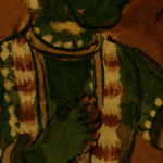
- Detail of 17th century Indian painting, ambient light. #1960.53

-
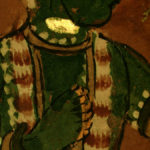
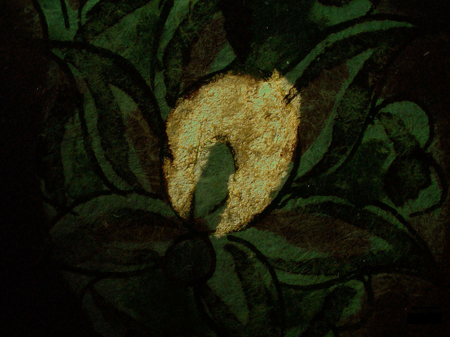
- Detail of 17th century Indian painting, ambient light plus fluorescence. #1960.53
-
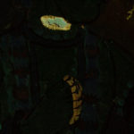
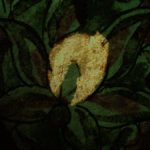
- Detail of 17th century Indian painting, fluorescence only. #1960.53
-
- Detail of 17th century Indian painting, ambient light. #1960.53
-
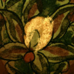
- Detail of 17th century Indian painting, ambient light plus fluorescence. #1960.53
-
- Detail of 17th century Indian painting, fluorescence only. #1960.53
-
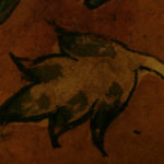
- Detail of 17th century Indian painting, ambient light. #1960.53

-
- Detail of 17th century Indian painting, ambient light plus fluorescence. #1960.53
-
- Detail of 17th century Indian painting, fluorescence only. #1960.53
-
- Detail of 17th century Indian painting, white light. #2006.262
-

- Detail of 17th century Indian painting, ambient light plus fluorescence. #2006.262
Subjects:
- Accession #1960.53. Krishna Receives Homage from a Prince: Possibly From a Bhagavata Purana Series. Ink, opaque watercolor and gold on paper. Unknown Artist, 17th century. Harvard Art Museums/Arthur M. Sackler Museum, Gift of John Goelet.
- Accession #2006.62. Malik Shah Rustam in the Presence of Shah Isma’il, illustrated folio from a manuscript of the Tarikh-i `Alamara-yi Shah Isma`il. Ink, opaque watercolor and gold on paper. Mu’in Musavvir, c. 1688. Harvard Art Museums/Arthur M. Sackler Museum, Gift of Mrs. Ezzat-Malek Soudavar in honor of Tom Lentz.
————
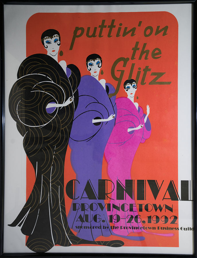
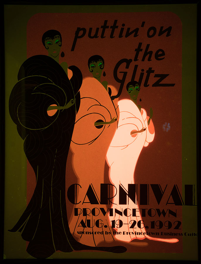
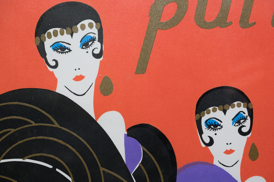
Poster print
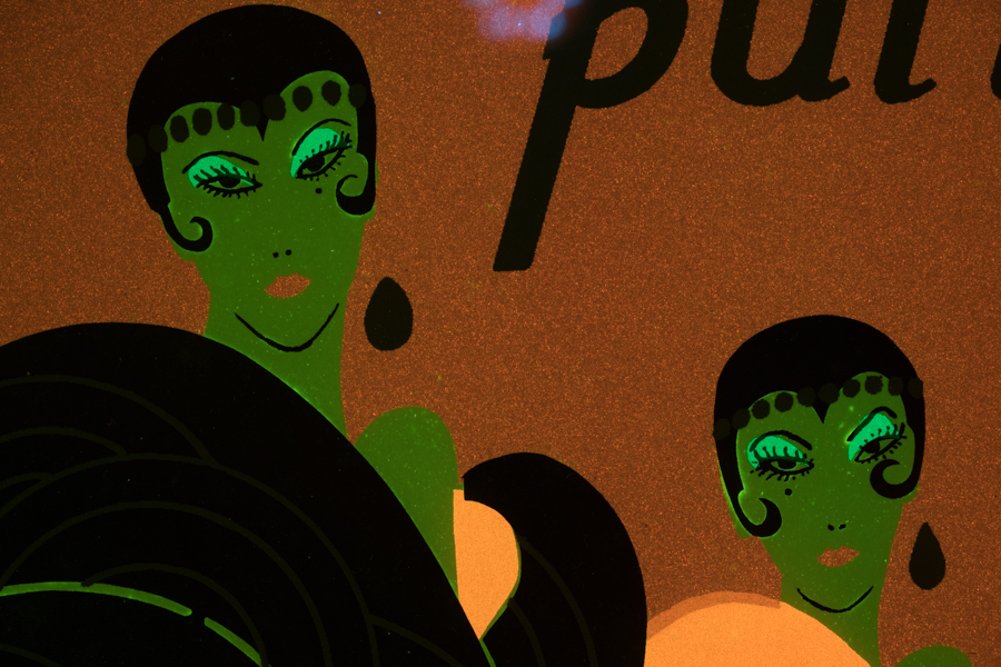
This example shows a poster made for the 1992 Carnival in Provincetown, MA. We found this print mounted on the wall of a B&B in Provincetown and photographed it with Royal Blue excitation.
-
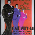
- Provincetown Carnival poster, white light.
-
- Provincetown Carnival poster, fluorescence.
-

- Provincetown Carnival poster detail, white light.
-
- Provincetown Carnival poster detail, fluorescence.
Touch-ups of a classic comic book cover
Fluorescence can potentially help to find areas of where restoration has been done. Someone sent us a classic comic book to see if fluorescence would show the touch-ups. As often happens fluorescence was not a universal panacea, and some areas did not show up particularly well. But in others fluorescence revealed the restorations dramatically. In some cases it was the touch-up itself that fluoresced, while in other cases it showed up dark against the natural fluorescence of the background.
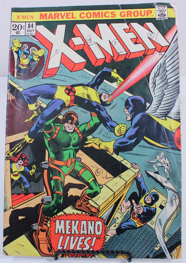
Cover of X-Men comic book from 1984
-
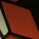
- Comic book cover detail, white light
-
- Comic book cover detail, fluorescence, Green excitation.
-
- Comic book cover detail, white light
-
- Comic book cover detail, fluorescence, Ultraviolet excitation.
-
- Comic book cover detail, white light
-
- Comic book cover detail, fluorescence, Royal Blue excitation.
Repairs
Fluorescence can aid in finding and documenting repairs. Belo
w are two examples we have come across:
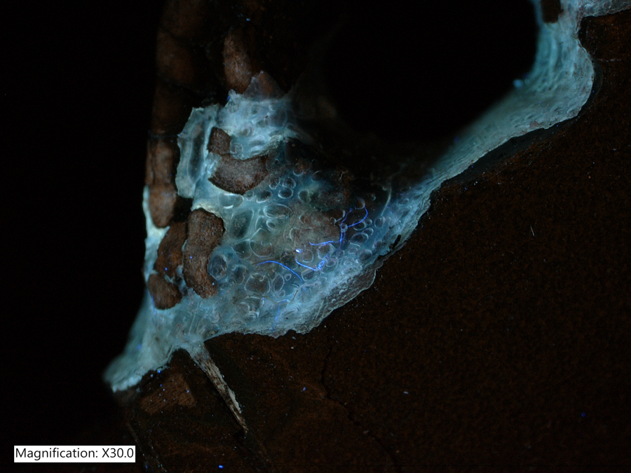
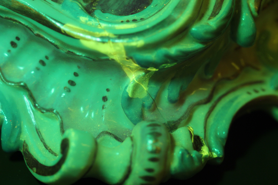
- Synthetic adhesive on Japanese lacquer pipe case (Kiseruzutsu). Image made with a Keyence VHX-6000 microscope with VH-Z20 lens, NIGHTSEA Ultraviolet excitation.
- Repair on a ceramic object. Macroscopic image, Royal Blue excitation.
-
- Synthetic adhesive on Japanese lacquer pipe case, Ultraviolet excitation
-
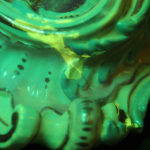
- Ceramic repair, Royal Blue excitation
Paper
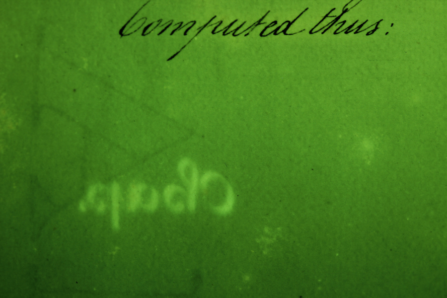
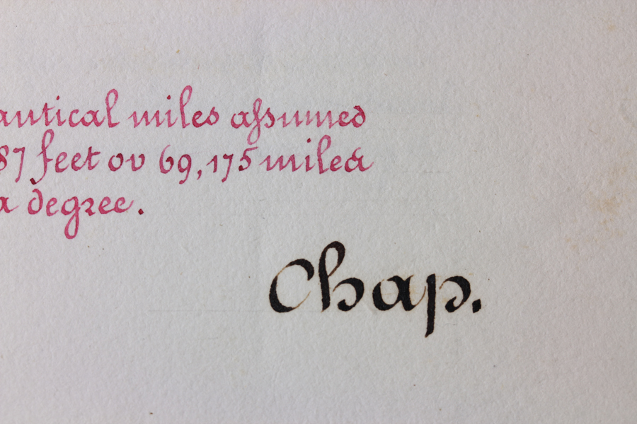
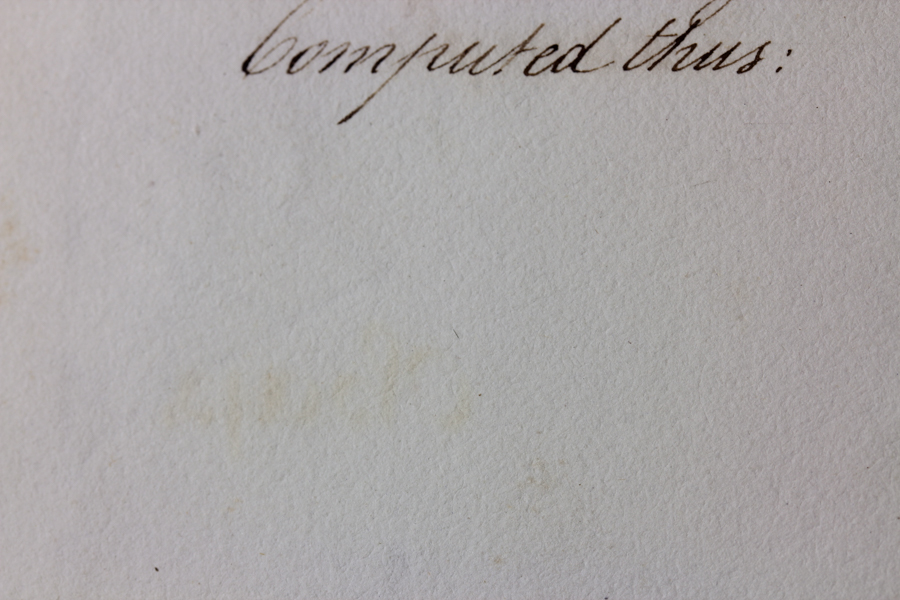
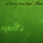
Fluorescence can reveal interesting features in paper. The images below were made of a c. 1800, beautifully bound and executed handwritten report on naval architecture experiments conducted in the 1790’s. We observed that all of the black ink could be seen on the facing sheet as an area of lighter fluorescence. According to an expert we consulted, this is due to transfer of unbound iron ions in the iron gall ink to the opposite page, where they form Iron (II) oxide. The fluorescence is not from the iron, but from chemical changes in the paper.
Image made with a macro lens on a DSLR camera. Fluorescence image made with Royal Blue excitation and a yellow barrier filter in front of the lens.
-
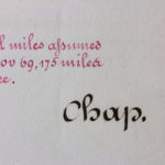
- Detail of c.1800 handwritten book, white light.
-
- Detail of c.1800 handwritten book, facing page, white light.
-
- Detail of c.1800 handwritten book, facing page, fluorescence.